Nahal Chadi, Wunker Claire, Keller Jennifer
Department of Surgery, Saint Louis University School of Medicine, St. Louis, MO, United States.
Front Surg. 2024 Nov 8;11:1413188. doi: 10.3389/fsurg.2024.1413188. eCollection 2024.
Papillary renal cell carcinoma accounts for one tenth of all renal cell carcinomas. Compared to other renal cell carcinoma subtypes, it is more often localized at the time of diagnosis and rarely metastasizes to the skin. There are no previously reported cases of cutaneous papillary renal cell carcinoma localized to the abdominal wall which we present here.
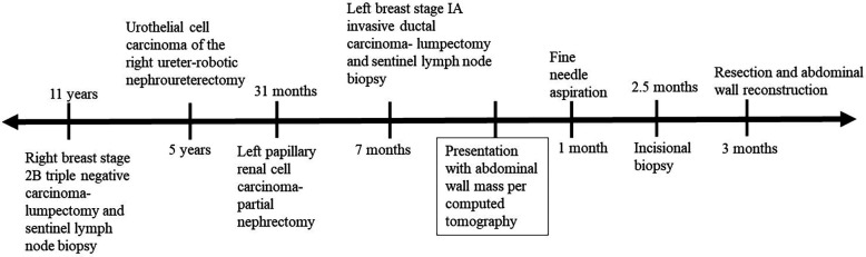
A 77 year-old female with multiple previous cancers, including a stage 1 left papillary renal cell carcinoma, treated with partial nephrectomy 32 months prior to presentation, was found to have a left upper abdominal wall mass on interval screening computed tomography. Fine needle aspiration was performed, obtaining limited tissue, followed by incisional biopsy. Histology and immunohistochemistry were consistent with renal cell carcinoma. She underwent operative excision of the abdominal wall mass with reconstruction using mesh and left posterior rectus fascial release. Histology and immunohistochemistry of the operative specimen reconfirmed the diagnosis of cutaneous metastasis of renal cell carcinoma. She was treated with adjuvant pembrolizumab and has no existing evidence of disease.
Papillary renal cell carcinoma metastasized to the skin is uncommon, especially when localized to the abdominal wall without any other sites of metastases. Metastasis should be on the differential diagnosis when evaluating newly identified abdominal masses in patients with a history of papillary renal cell carcinoma. When localized, abdominal wall metastasis of papillary renal cell carcinoma can be effectively treated with resection and reconstruction, followed by systemic therapy when indicated.
乳头状肾细胞癌占所有肾细胞癌的十分之一。与其他肾细胞癌亚型相比,它在诊断时更常局限,很少转移至皮肤。此前尚无局限于腹壁的皮肤乳头状肾细胞癌的病例报道,我们在此呈现。
一名77岁女性,既往有多种癌症,包括1期左乳头状肾细胞癌,在就诊前32个月接受了部分肾切除术,在定期筛查计算机断层扫描时发现左上腹壁有一肿块。进行了细针穿刺抽吸,获取的组织有限,随后进行了切开活检。组织学和免疫组化结果与肾细胞癌一致。她接受了腹壁肿块的手术切除,并用网片进行重建以及左腹直肌后鞘松解。手术标本的组织学和免疫组化再次证实为肾细胞癌皮肤转移。她接受了辅助派姆单抗治疗,目前没有疾病迹象。
乳头状肾细胞癌转移至皮肤并不常见,尤其是局限于腹壁且无其他转移部位时。在评估有乳头状肾细胞癌病史患者新发现的腹部肿块时,应将转移纳入鉴别诊断。当局限时,乳头状肾细胞癌腹壁转移可通过切除和重建有效治疗,必要时随后进行全身治疗。