In situ Structural Biology, Max Delbrück Center for Molecular Medicine in the Helmholtz Association (MDC), Berlin 13125, Germany.
Department of Biology, Humboldt University of Berlin, Berlin, Germany.
Proc Natl Acad Sci U S A. 2024 Dec 3;121(49):e2407375121. doi: 10.1073/pnas.2407375121. Epub 2024 Nov 27.
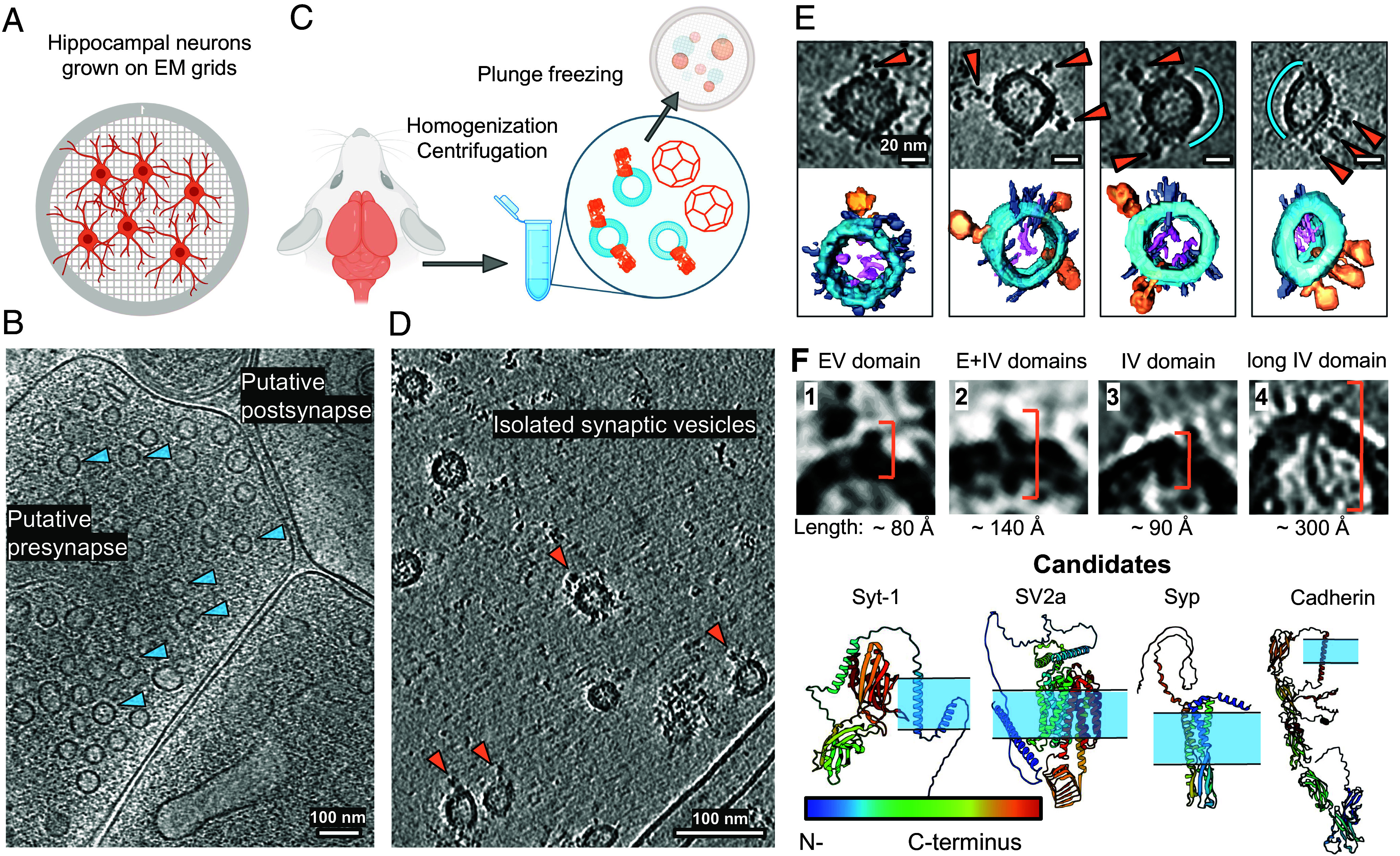
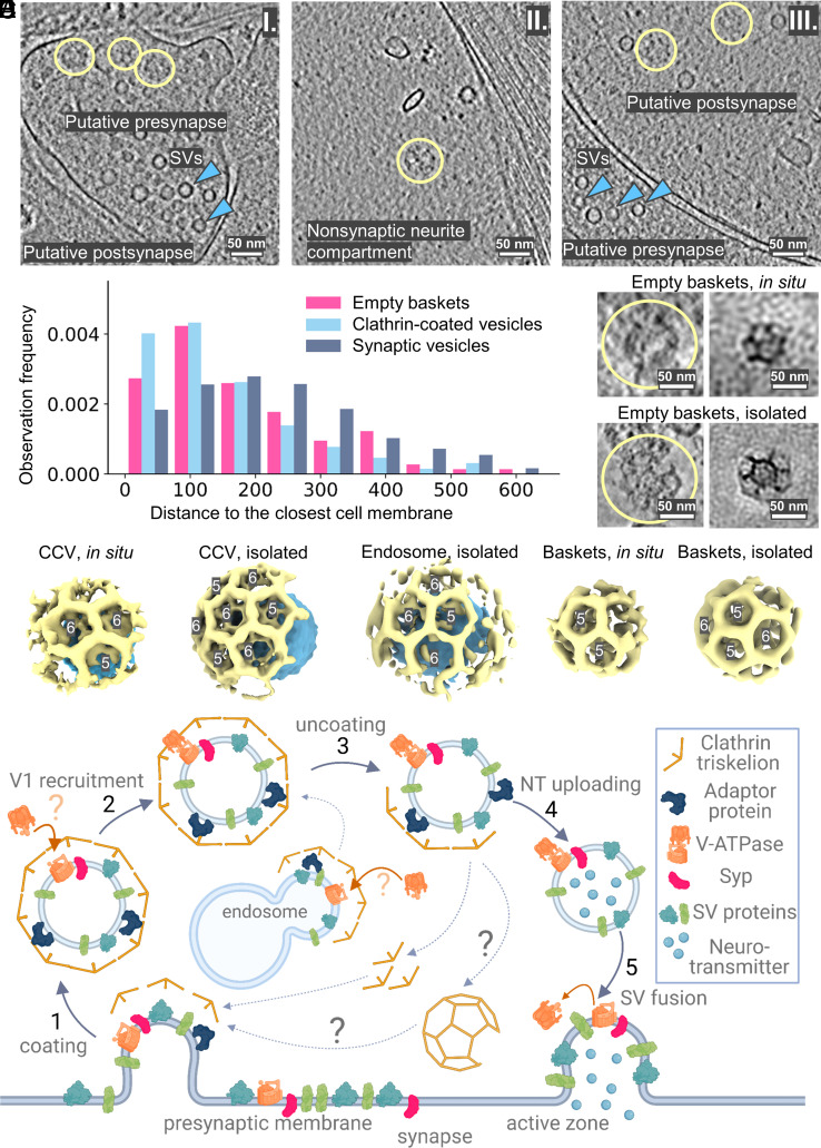
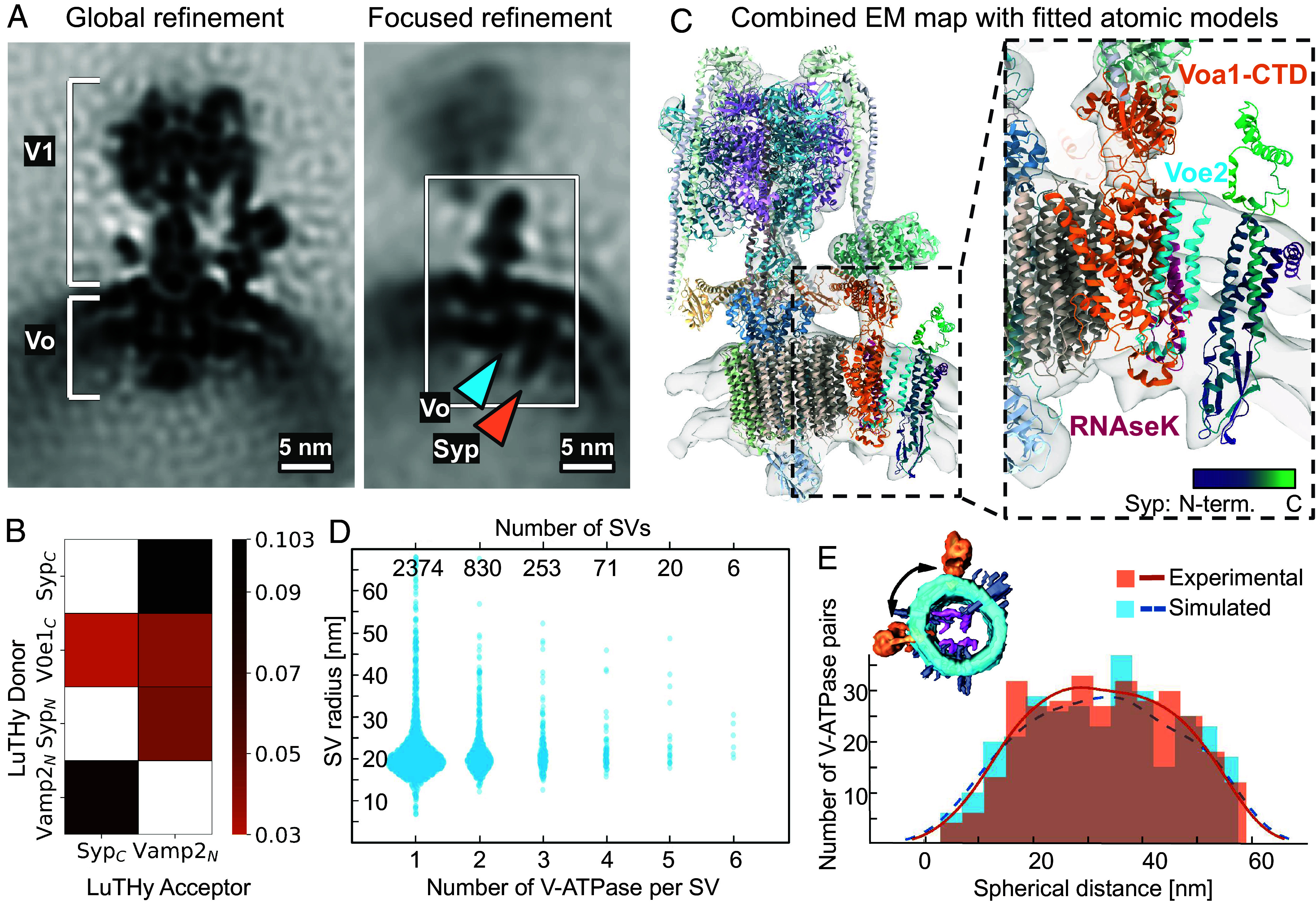
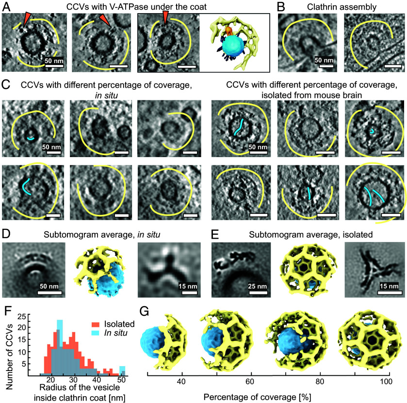
Synaptic vesicles (SVs) store and transport neurotransmitters to the presynaptic active zone for release by exocytosis. After release, SV proteins and excess membrane are recycled via endocytosis, and new SVs can be formed in a clathrin-dependent manner. This process maintains complex molecular composition of SVs through multiple recycling rounds. Previous studies explored the molecular composition of SVs through proteomic analysis and fluorescent microscopy, proposing a model for an average SV (1). However, the structural heterogeneity and molecular architecture of individual SVs are not well described. Here, we used cryoelectron tomography to visualize molecular details of SVs isolated from mouse brains and inside cultured neurons. We describe several classes of small proteins on the SV surface and long proteinaceous densities inside SVs. We identified V-ATPases, determined a structure using subtomogram averaging, and showed them forming a complex with the membrane-embedded protein synaptophysin (Syp). Our bioluminescence assay revealed pairwise interactions between vesicle-associated membrane protein 2 and Syp and V-ATPase Voe1 domains. Interestingly, V-ATPases were randomly distributed on the surface of SVs irrespective of vesicle size. A subpopulation of isolated vesicles and vesicles inside neurons contained a partially assembled clathrin coat with an icosahedral symmetry. We observed V-ATPases under clathrin cages in several isolated clathrin-coated vesicles (CCVs). Additionally, from isolated SV preparations and within hippocampal neurons we identified clathrin baskets without vesicles. We determined their and CCVs preferential location in proximity to the cell membrane. Our analysis advances the understanding of individual SVs' diversity and their molecular architecture.
突触小泡 (SVs) 储存并将神经递质运输到突触前活性区,通过胞吐作用释放。释放后,SV 蛋白和多余的膜通过胞吞作用被回收,新的 SV 可以以网格蛋白依赖的方式形成。这个过程通过多个回收循环来维持 SV 的复杂分子组成。先前的研究通过蛋白质组学分析和荧光显微镜探索了 SV 的分子组成,提出了一个平均 SV 的模型 (1)。然而,单个 SV 的结构异质性和分子结构尚未得到很好的描述。在这里,我们使用冷冻电子断层扫描来可视化从小鼠大脑中分离出的 SV 以及培养神经元内的 SV 的分子细节。我们描述了 SV 表面上的几类小蛋白和 SV 内部的长蛋白密度。我们鉴定了 V-ATPases,使用亚体素平均法确定了结构,并显示它们与膜嵌入蛋白突触小泡蛋白 (Syp) 形成复合物。我们的生物发光测定揭示了囊泡相关膜蛋白 2 和 Syp 与 V-ATPase Voe1 结构域之间的成对相互作用。有趣的是,V-ATPases在 SV 表面上的分布是随机的,与囊泡大小无关。分离的囊泡和神经元内的囊泡亚群包含具有二十面体对称性的部分组装的网格蛋白涂层。我们在几个分离的网格蛋白包被囊泡 (CCVs) 中观察到 V-ATPases 位于网格蛋白笼下。此外,我们从分离的 SV 制剂和海马神经元中鉴定出没有囊泡的网格蛋白篮。我们确定了它们和 CCVs 在靠近细胞膜的位置的优先位置。我们的分析推进了对单个 SV 多样性及其分子结构的理解。