Roche Pharma Research and Early Development, Neuroscience and Rare Diseases, Roche Innovation Center Basel, F.Hoffmann-La Roche Ltd, Basel, Switzerland.
Ace Alzheimer Center Barcelona - Universitat Internacional de Catalunya, Barcelona, Spain.
Alzheimers Res Ther. 2024 Nov 28;16(1):257. doi: 10.1186/s13195-024-01622-5.
Second-generation tau tracers for positron emission tomography (PET) show high affinity for paired helical filaments tau deposits characteristic of Alzheimer´s disease and low off-target binding. Differences in their chemical structure though may lead to variations in their regional tau uptake and off-target signal. In this work, we aimed to compare the in-vivo uptake of tau tracers [F]PI-2620 and [F]RO948 in the early stages of the AD continuum.
Data from the TAU-PET FACEHBI clinical trial (EUDRA-CT 2021-000473-83) were analyzed. All participants were non-demented and underwent tau imaging with [F]PI-2620 and [F]RO948 PET within 3 months, amyloid imaging with [F]Florbetaben and brain magnetic resonance imaging. Tau PET standardized uptake values ratios (SUVR) were calculated in Braak and typical off-target regions using the inferior cerebellar cortex as a reference region.
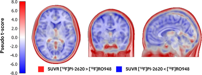
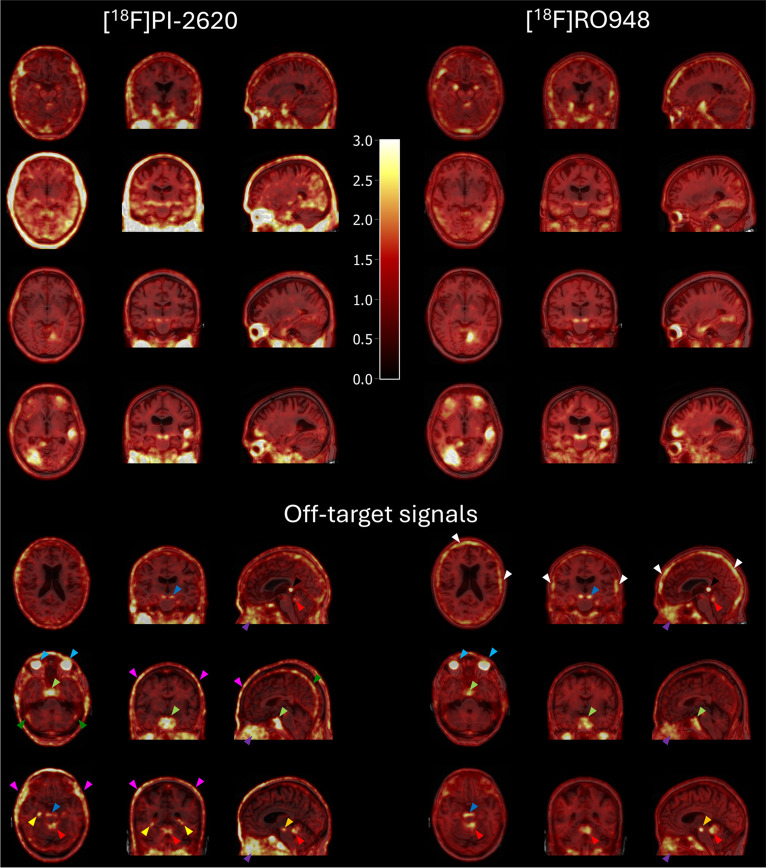
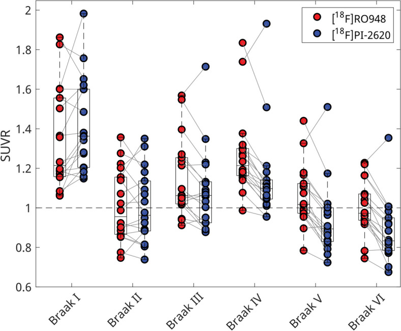
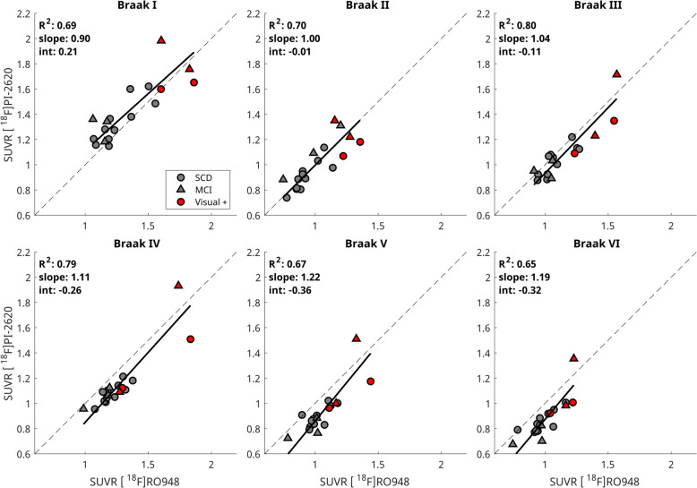
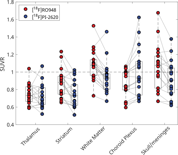
The cohort consisted of 18 individuals with subjective cognitive decline (n = 13) and mild cognitive impairment (n = 5), with centiloid values ranging from 17 to 159. Both tau tracers showed similar tau pathology distribution but presented a distinct off-target signal pattern on visual read. SUVR measurements for [F]PI-2620 and [F]RO948 were highly correlated in all Braak regions (R range [0.65-0.80]). Regarding off-target signal, [F]PI-2620 had higher SUVRs in vascular structures, and [F]RO948 had higher SUVRs in the skull/meninges.
In a cohort of individuals at early stages of the AD continuum, tau PET tracers [F]PI-2620 and [F]RO948 showed similar in-vivo uptake in all Braak regions and distinct off-target signal. These preliminary results support the development of standardized quantification scales for tau deposition that are tracer-independent.
AEMPS EudraCT 2021-000473-83. Registered 30 December 2021.
正电子发射断层扫描(PET)的第二代 tau 示踪剂对阿尔茨海默病特征性的双螺旋细丝 tau 沉积物具有高亲和力和低脱靶结合。然而,它们的化学结构差异可能导致其在 tau 摄取和脱靶信号方面的差异。在这项工作中,我们旨在比较 AD 连续体早期阶段 tau 示踪剂[F]PI-2620 和[F]RO948 的体内摄取。
分析了 TAU-PET FACEHBI 临床试验(EUDRA-CT 2021-000473-83)的数据。所有参与者均无痴呆,并在 3 个月内接受 tau 成像[F]PI-2620 和[F]RO948 PET、[F]Florbetaben 淀粉样蛋白成像和脑磁共振成像。tau PET 标准化摄取值比(SUVR)在 Braak 和典型脱靶区域使用小脑下蚓作为参考区域进行计算。
该队列包括 18 名有主观认知减退(n=13)和轻度认知障碍(n=5)的个体,百分位数值范围为 17 至 159。两种 tau 示踪剂均显示出相似的 tau 病理学分布,但在视觉阅读中表现出不同的脱靶信号模式。[F]PI-2620 和[F]RO948 的 SUVR 测量值在所有 Braak 区域均高度相关(R 范围为[0.65-0.80])。关于脱靶信号,[F]PI-2620 在血管结构中具有更高的 SUVR,[F]RO948 在颅骨/脑膜中具有更高的 SUVR。
在 AD 连续体早期阶段的个体队列中,tau PET 示踪剂[F]PI-2620 和[F]RO948 在所有 Braak 区域的体内摄取相似,脱靶信号不同。这些初步结果支持开发tau 沉积的标准化定量量表,该量表与示踪剂无关。
AEMPS EudraCT 2021-000473-83。于 2021 年 12 月 30 日注册。