Peretzke Robin, Neher Peter F, Brandt Geva A, Fritze Stefan, Volkmer Sebastian, Daub Jonas, Northoff Georg, Bohn Jonas, Kirchhoff Yannick, Roy Saikat, Maier-Hein Klaus H, Meyer-Lindenberg Andreas, Hirjak Dusan
Division of Medical Image Computing, German Cancer Research Center, Heidelberg, Germany.
Medical Faculty, Heidelberg University, Heidelberg, Germany.
Mol Psychiatry. 2025 May;30(5):2095-2107. doi: 10.1038/s41380-024-02821-0. Epub 2024 Dec 2.
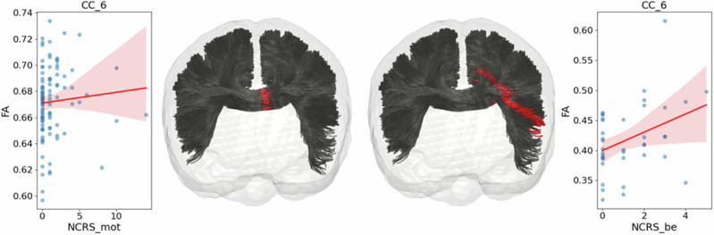
Catatonia is a severe psychomotor disorder characterized by motor, affective and cognitive-behavioral abnormalities. Although previous magnetic resonance imaging (MRI) studies suggested white matter (WM) dysconnectivity in the pathogenesis of catatonia, it is unclear whether microstructural alterations of WM tracts connecting psychomotor regions might contribute to a better classification of catatonia patients. Here, diffusion-weighted MRI data were collected from two independent cohorts (whiteCAT/replication cohort) of patients with (n = 45/n = 13) and without (n = 56/n = 26) catatonia according to ICD-11 criteria. Catatonia severity was examined using the Northoff (NCRS) and Bush-Francis (BFCRS) Catatonia Rating Scales. We used tract-based spatial statistics (TBSS), tractometry (TractSeg) and machine-learning (ML) to classify catatonia patients from tractometry values as well as tractomics features generated by the newly developed tool RadTract. Catatonia patients showed fractional anisotropy (FA) alterations measured via TractSeg in different corpus callosum segments (CC_1, CC_3, CC_4, CC_5 and CC_6) compared to non-catatonia patients across both cohorts. Our classification results indicated a higher level of performance when trained on tractomics as opposed to traditional tractometry values. Moreover, in the CC_6, we successfully trained two classifiers using the tractomics features identified in the whiteCAT data. These classifiers were applied separately to the whiteCAT and replication cohorts, demonstrating comparable performance with Area Under the Receiver Operating Characteristics (AUROC) values of 0.79 for the whiteCAT cohort and 0.76 for the replication cohort. In contrast, training on FA tractometry resulted in lower AUROC values of 0.66 for the whiteCAT cohort and 0.51 for the replication cohort. In conclusion, these findings underscore the significance of CC WM microstructural alterations in the pathophysiology of catatonia. The successful use of an ML based classification model to identify catatonia patients has the potential to improve diagnostic precision.
紧张症是一种严重的精神运动障碍,其特征为运动、情感及认知行为异常。尽管先前的磁共振成像(MRI)研究提示白质(WM)连接障碍在紧张症发病机制中起作用,但连接精神运动区域的WM束的微观结构改变是否有助于更好地对紧张症患者进行分类尚不清楚。在此,根据国际疾病分类第11版(ICD-11)标准,从两个独立队列(whiteCAT/复制队列)中收集了弥散加权MRI数据,其中一组为紧张症患者(n = 45/n = 13),另一组为非紧张症患者(n = 56/n = 26)。使用诺托夫(NCRS)和布什-弗朗西斯(BFCRS)紧张症评定量表检查紧张症严重程度。我们使用基于束的空间统计学(TBSS)、束测量法(TractSeg)和机器学习(ML),根据束测量值以及新开发工具RadTract生成的束组学特征对紧张症患者进行分类。与两个队列中的非紧张症患者相比,紧张症患者在不同胼胝体节段(CC_1、CC_3、CC_4、CC_5和CC_6)通过TractSeg测量的各向异性分数(FA)发生了改变。我们的分类结果表明,与传统的束测量值相比,基于束组学进行训练时的性能水平更高。此外,在CC_6中,我们使用在whiteCAT数据中识别出的束组学特征成功训练了两个分类器。这些分类器分别应用于whiteCAT和复制队列,在whiteCAT队列中的受试者工作特征曲线下面积(AUROC)值为0.79,在复制队列中的AUROC值为0.76,表现相当。相比之下,基于FA束测量进行训练时,whiteCAT队列的AUROC值为0.66,复制队列的AUROC值为0.51,较低。总之,这些发现强调了CC白质微观结构改变在紧张症病理生理学中的重要性。成功使用基于ML的分类模型识别紧张症患者有可能提高诊断准确性。