Hoffmann Tobias, Michel Janine, Nitsche Andreas, Mache Christin, Schulze Jessica, Wolff Thorsten, Laue Michael
Advanced Light and Electron Microscopy, Centre for Biological Threats and Special Pathogens 4 (ZBS 4), Robert Koch Institute, Berlin, Germany.
Highly Pathogenic Viruses, Centre for Biological Threats and Special Pathogens 1 (ZBS 1), Robert Koch Institute, Berlin, Germany.
Sci Data. 2024 Dec 4;11(1):1322. doi: 10.1038/s41597-024-04182-3.
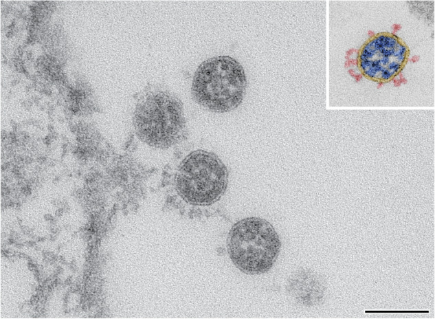
Conventional thin section electron microscopy of viral pathogens, such as the pandemic SARS-CoV-2, can provide structural information on the virus particle phenotype and its evolution. We recorded about 900 transmission electron microscopy images of different SARS-CoV-2 variants, including Alpha (B.1.1.7), Beta (B.1.351), Delta (B.1.617.2) and Omicron BA.2 (B.1.1.529) and determined various morphometric parameters, such as maximal diameter and spike number, using a previously published measurement method. The datasets of the evolved virus variants were supplemented with images and measurements of the early SARS-CoV-2 isolates Munich929 and Italy-INMI1 to allow direct comparison. Infected Vero cell cultures were cultivated under comparable conditions to produce the viruses for imaging and morphometric analysis. The images and measurements can be used as a basis to analyse the morphometric changes of further evolving viruses at the particle level or for developing automated image processing workflows and analysis.
对诸如大流行的严重急性呼吸综合征冠状病毒2(SARS-CoV-2)等病毒病原体进行传统的超薄切片电子显微镜检查,可以提供有关病毒颗粒表型及其进化的结构信息。我们记录了约900张不同SARS-CoV-2变体的透射电子显微镜图像,包括阿尔法(B.1.1.7)、贝塔(B.1.351)、德尔塔(B.1.617.2)和奥密克戎BA.2(B.1.1.529),并使用先前发表的测量方法确定了各种形态测量参数,如最大直径和刺突数量。进化后的病毒变体数据集补充了早期SARS-CoV-2分离株慕尼黑929和意大利-INMI1的图像和测量数据,以便进行直接比较。在可比条件下培养受感染的Vero细胞培养物,以生产用于成像和形态测量分析的病毒。这些图像和测量数据可作为在颗粒水平分析进一步进化的病毒的形态变化的基础,或用于开发自动化图像处理工作流程和分析。