Yin Li, Liao Dongfa, Xie Qingyun, Liu Jinbiao, Deng Bing
Department of Orthopaedics, General Hospital of Western Theater Command, 270 Tianhui Rd, Chengdu, Sichuan, PR China.
J Orthop Surg Res. 2024 Dec 4;19(1):822. doi: 10.1186/s13018-024-05306-6.
To investigate the anatomical features of the femoral tunnel in anatomical and isometric single-bundle ACL reconstruction.
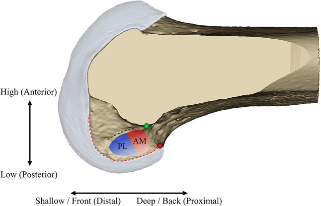
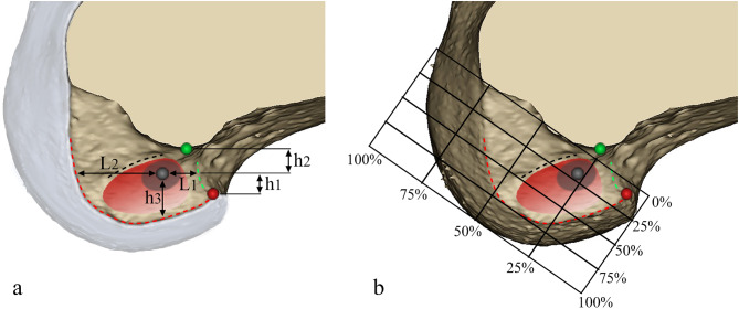
Thirty-two 3-dimensional knee models were reconstructed based on CT scan (average age: 26.5 ± 6.7 years, 18 males and 14 females, 17 left and 15 right). Multiple anatomical landmarks were identified. Virtual femoral tunnels were created at the deep and high portion of ACL footprint, close to the lateral intercondylar ridge to achieve best anatomy and isometry, simulating an anteromedial portal reconstruction. Anatomical features of the femoral tunnels were analyzed. The position of the femoral tunnel was quantified by the distance to anatomical landmarks and using quadrant methods. The spatial angles, length and outer opening of the femoral tunnels were also evaluated.
Acceptable tunnels were created in all models. The center of femoral tunnel was slightly higher than the apex of deep cartilage, near the deep one-third point across the shallow-deep dimension of the lateral femoral condyle. Using the quadrant method, the tunnel was located at 28.4% ± 2.2% and 22.2% ± 3.6%, parallel and perpendicular to the Blumensaat line, respectively. The spatial angles of the tunnel were 40°, 33.5° ± 4.1° and 38.2° ± 4.4° on the sagittal, transverse, and coronal planes, respectively. The average tunnel length was 34.8 mm ± 3.8 mm. The outer opening of the tunnels was located at the posterior one-third of the femoral metaphysis.
The anatomical and isometric positioning of the femoral tunnel can be achieved through anteromedial portal with satisfied tunnel characteristics. The apex of deep cartilage may be used as an anatomical reference for tunnel positioning. When drilled at appropriate orientation, favorable tunnel length, integrity and position of the outer opening can be obtained.
研究解剖学单束和等距单束前交叉韧带(ACL)重建中股骨隧道的解剖学特征。
基于CT扫描重建32个三维膝关节模型(平均年龄:26.5±6.7岁,男性18例,女性14例,左侧17例,右侧15例)。确定多个解剖标志。在ACL足迹的深部和高处、靠近外侧髁间嵴处创建虚拟股骨隧道,以实现最佳解剖结构和等距性,模拟前内侧入路重建。分析股骨隧道的解剖学特征。通过与解剖标志的距离和象限法对股骨隧道的位置进行量化。还评估了股骨隧道的空间角度、长度和外口。
所有模型均创建出可接受的隧道。股骨隧道中心略高于深层软骨顶点,靠近股骨外侧髁浅深维度的后三分之一处。采用象限法,隧道分别位于与布卢门萨特线平行和垂直方向的28.4%±2.2%和22.2%±3.6%处。隧道在矢状面、横断面和冠状面的空间角度分别为40°、33.5°±4.1°和38.2°±4.4°。平均隧道长度为34.8 mm±3.8 mm。隧道外口位于股骨干骺端的后三分之一处。
通过前内侧入路可实现股骨隧道的解剖学和等距定位,且隧道特征良好。深层软骨顶点可作为隧道定位的解剖学参考。以适当方向钻孔时,可获得理想的隧道长度、完整性和外口位置。