Wang Hao, He Limeng, Feng Lijuan, Zhang Weiwei, Liu Nan, Zhang Wei
Department of Nuclear Medicine, Sichuan Provincial People's Hospital, University of Electronic Science and Technology of China, Chengdu Sichuan, China.
Clin Kidney J. 2024 Nov 11;17(12):sfae340. doi: 10.1093/ckj/sfae340. eCollection 2024 Dec.
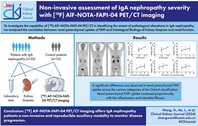
Renal biopsy plays a crucial role in diagnosing and assessing the severity of immunoglobulin A nephropathy (IgAN), despite being an invasive procedure with potential risk of failure. Our study focused on evaluating the capability of [F]AlF-NOTA-FAPI-04 PET/CT in identifying the extent of pathological alterations in IgAN.
Twenty patients (13 males and 7 females; mean age, 44 ± 16 years) with newly diagnosed primary IgAN and 10 patients (7 males and 3 females; mean age, 51 ± 4 years) without known renal disease underwent [F]AlF-NOTA-FAPI-04 PET/CT imaging. Kidney tissues from biopsies were stained with various techniques and examined using immunofluorescence. The Oxford classification was used to evaluate pathological indicators. Immunohistochemical staining was conducted to assess α-smooth muscle actin (αSMA) and fibroblast activation protein (FAP) expression. Renal FAPI uptake measured by positron emission tomography/computed tomography (PET/CT) (maximum and mean standardized uptake value, SUV and SUV) was correlated with histological findings.
The renal parenchymal FAPI uptake was significantly higher in IgAN patients compared with control patients (SUV= 3.9 ± 1.3 vs 1.9 ± 0.4, SUV = 3.6 ± 1.2 vs 1.5 ± 0.4; all < .001). We identified a significant difference in renal parenchymal FAPI uptake among the various categories of the Oxford classification. Correlation analysis revealed a positive association between SUV and interstitial fibrosis and tubular atrophy, as well as tubulointerstitial inflammation scores in scarred cortex and non-scarred cortex ( = 0.637, 0.593 and 0.491, all < .05), Similar associations were observed between SUV and these scores ( = 0.641, 0.592 and 0.479, all < .05). Furthermore, significant positive correlations were observed between SUV or SUV and the staining scores for glomerular αSMA and FAP, as well as for tubulointerstitial αSMA and FAP (all < .01).
[F]AlF-NOTA-FAPI-04 PET/CT imaging offers IgAN patients a non-invasive and reproducible auxiliary modality to monitor disease progression.
肾活检在免疫球蛋白A肾病(IgAN)的诊断和严重程度评估中起着关键作用,尽管它是一种具有潜在失败风险的侵入性检查。我们的研究聚焦于评估[F]AlF-NOTA-FAPI-04 PET/CT识别IgAN病理改变程度的能力。
20例新诊断的原发性IgAN患者(13例男性,7例女性;平均年龄44±16岁)和10例无已知肾脏疾病的患者(7例男性,3例女性;平均年龄51±4岁)接受了[F]AlF-NOTA-FAPI-04 PET/CT成像。活检获取的肾组织采用多种技术染色,并进行免疫荧光检查。采用牛津分类法评估病理指标。进行免疫组织化学染色以评估α平滑肌肌动蛋白(αSMA)和成纤维细胞活化蛋白(FAP)的表达。通过正电子发射断层扫描/计算机断层扫描(PET/CT)测量的肾脏FAPI摄取(最大和平均标准化摄取值,SUV和SUV)与组织学结果相关。
与对照组患者相比,IgAN患者的肾实质FAPI摄取显著更高(SUV=3.9±1.3 vs 1.9±0.4,SUV=3.6±1.2 vs 1.5±0.4;均P<0.001)。我们在牛津分类的不同类别中发现肾实质FAPI摄取存在显著差异。相关性分析显示,SUV与间质纤维化、肾小管萎缩以及瘢痕皮质和非瘢痕皮质的肾小管间质炎症评分之间呈正相关(r=0.637、0.593和0.491,均P<0.05),在SUV与这些评分之间也观察到类似的相关性(r=0.641、0.592和0.479,均P<0.05)。此外,在SUV或SUV与肾小球αSMA和FAP以及肾小管间质αSMA和FAP的染色评分之间观察到显著的正相关(均P<0.01)。
[F]AlF-NOTA-FAPI-04 PET/CT成像为IgAN患者提供了一种无创且可重复的辅助手段来监测疾病进展。