Department of Radiology and Nuclear Medicine, University Medical Center Mannheim, Heidelberg University, Theodor-Kutzer-Ufer 1-3, 68167, Mannheim, Germany.
Department of Radiology, German Cancer Research Center, Im Neuenheimer Feld 230, 69120, Heidelberg, Germany.
Sci Rep. 2022 Nov 15;12(1):19594. doi: 10.1038/s41598-022-22877-8.
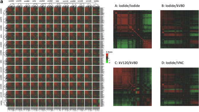
Feature stability and standardization remain challenges that impede the clinical implementation of radiomics. This study investigates the potential of spectral reconstructions from photon-counting computed tomography (PCCT) regarding organ-specific radiomics feature stability. Abdominal portal-venous phase PCCT scans of 10 patients in virtual monoenergetic (VM) (keV 40-120 in steps of 10), polyenergetic, virtual non-contrast (VNC), and iodine maps were acquired. Two 2D and 3D segmentations measuring 1 and 2 cm in diameter of the liver, lung, spleen, psoas muscle, subcutaneous fat, and air were obtained for spectral reconstructions. Radiomics features were extracted with pyradiomics. The calculation of feature-specific intraclass correlation coefficients (ICC) was performed by comparing all segmentation approaches and organs. Feature-wise and organ-wise correlations were evaluated. Segmentation-resegmentation stability was evaluated by concordance correlation coefficient (CCC). Compared to non-VM, VM-reconstruction features tended to be more stable. For VM reconstructions, 3D 2 cm segmentation showed the highest average ICC with 0.63. Based on a criterion of ≥ 3 stable organs and an ICC of ≥ 0.75, 12-mainly non-first-order features-are shown to be stable between the VM reconstructions. In a segmentation-resegmentation analysis in 3D 2 cm, three features were identified as stable based on a CCC of > 0.6 in ≥ 3 organs in ≥ 6 VM reconstructions. Certain radiomics features vary between monoenergetic reconstructions and depend on the ROI size. Feature stability was also shown to differ between different organs. Yet, glcm_JointEntropy, gldm_GrayLevelNonUniformity, and firstorder_Entropy could be identified as features that could be interpreted as energy-independent and segmentation-resegmentation stable in this PCCT collective. PCCT may support radiomics feature standardization and comparability between sites.
特征稳定性和标准化仍然是阻碍放射组学临床应用的挑战。本研究探讨了基于能谱光子计数 CT(PCCT)的器官特异性放射组学特征稳定性的光谱重建的潜力。对 10 名患者进行腹部门静脉期 PCCT 扫描,在虚拟单能量(VM)(keV 40-120,每 10keV 一步)、多能量、虚拟非对比(VNC)和碘图下进行。对肝脏、肺、脾脏、腰大肌、皮下脂肪和空气进行了直径为 1cm 和 2cm 的 2D 和 3D 分割,以进行光谱重建。使用 pyradiomics 提取放射组学特征。通过比较所有分割方法和器官,计算特征特定的组内相关系数(ICC)。评估特征和器官的相关性。通过一致性相关系数(CCC)评估分割再分割稳定性。与非 VM 相比,VM 重建特征的稳定性更高。对于 VM 重建,3D 2cm 分割的平均 ICC 最高,为 0.63。根据≥3 个稳定器官和 ICC≥0.75的标准,12 个主要非一级特征在 VM 重建之间被证明是稳定的。在 3D 2cm 的分割再分割分析中,根据 CCC>0.6 的标准,在≥3 个 VM 重建中,有 3 个特征被确定为稳定。某些放射组学特征在单能量重建之间存在差异,并且取决于 ROI 大小。特征稳定性在不同器官之间也存在差异。然而,glcm_JointEntropy、gldm_GrayLevelNonUniformity 和 firstorder_Entropy 可以被识别为在这个 PCCT 集合中可以被解释为能量独立和分割再分割稳定的特征。PCCT 可能支持放射组学特征的标准化和不同站点之间的可比性。