Department of Radiology and Nuclear Medicine, University Medical Center Mannheim, Heidelberg University, Theodor-Kutzer-Ufer 1-3, 68167, Mannheim, Germany.
Department of Radiology, German Cancer Research Center, Im Neuenheimer Feld 280, 69120, Heidelberg, Germany.
Eur Radiol. 2023 Jul;33(7):4905-4914. doi: 10.1007/s00330-023-09460-z. Epub 2023 Feb 21.
Radiomics image data analysis offers promising approaches in research but has not been implemented in clinical practice yet, partly due to the instability of many parameters. The aim of this study is to evaluate the stability of radiomics analysis on phantom scans with photon-counting detector CT (PCCT).
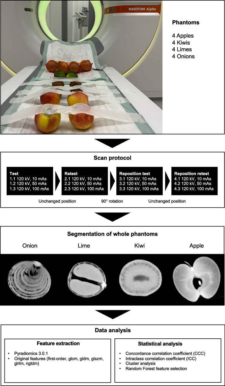
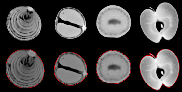
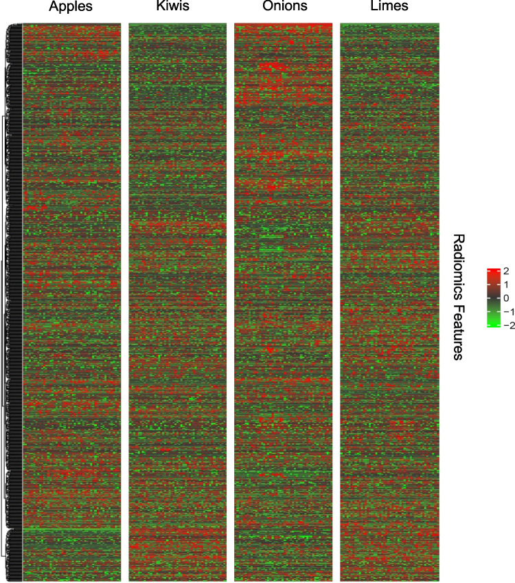
Photon-counting CT scans of organic phantoms consisting of 4 apples, kiwis, limes, and onions each were performed at 10 mAs, 50 mAs, and 100 mAs with 120-kV tube current. The phantoms were segmented semi-automatically and original radiomics parameters were extracted. This was followed by statistical analysis including concordance correlation coefficients (CCC), intraclass correlation coefficients (ICC), as well as random forest (RF) analysis, and cluster analysis to determine the stable and important parameters.
Seventy-three of the 104 (70%) extracted features showed excellent stability with a CCC value > 0.9 when compared in a test and retest analysis, and 68 features (65.4%) were stable compared to the original in a rescan after repositioning. Between the test scans with different mAs values, 78 (75%) features were rated with excellent stability. Eight radiomics features were identified that had an ICC value greater than 0.75 in at least 3 of 4 groups when comparing the different phantoms in a phantom group. In addition, the RF analysis identified many features that are important for distinguishing the phantom groups.
Radiomics analysis using PCCT data provides high feature stability on organic phantoms, which may facilitate the implementation of radiomics analysis likewise in clinical routine.
• Radiomics analysis using photon-counting computed tomography provides high feature stability. • Photon-counting computed tomography may pave the way for implementation of radiomics analysis in clinical routine.
放射组学图像数据分析在研究中提供了有前景的方法,但尚未在临床实践中实施,部分原因是许多参数不稳定。本研究旨在评估基于光子计数探测器 CT(PCCT)的体模扫描的放射组学分析的稳定性。
对由 4 个苹果、猕猴桃、酸橙和洋葱组成的有机体模进行光子计数 CT 扫描,管电流为 120 kV 时分别使用 10 mAs、50 mAs 和 100 mAs。使用半自动分割方法对体模进行分割,并提取原始放射组学参数。随后进行统计分析,包括一致性相关系数(CCC)、组内相关系数(ICC)、随机森林(RF)分析和聚类分析,以确定稳定和重要的参数。
在测试和复测分析中,73 个(70%)提取的特征的 CCC 值>0.9,表明具有极好的稳定性,68 个特征(65.4%)在重新定位后的重新扫描中与原始特征稳定。在不同 mAs 值的测试扫描中,78 个(75%)特征被评为极好的稳定性。当在体模组中比较不同的体模时,有 8 个放射组学特征在至少 3 个体模中被鉴定为 ICC 值大于 0.75。此外,RF 分析确定了许多对于区分体模组很重要的特征。
使用 PCCT 数据进行放射组学分析在有机体模上提供了高特征稳定性,这可能同样有助于在临床常规中实施放射组学分析。
• 使用光子计数计算机断层扫描的放射组学分析提供了高特征稳定性。
• 光子计数计算机断层扫描可能为在临床常规中实施放射组学分析铺平道路。