Xia Fei, Sinefeld David, Chang Zong, Gong Xiaojing, Sun Qinchao
School of Applied and Engineering Physics, Cornell University, Ithaca, New York, USA.
Meinig School of Biomedical Engineering, Cornell University, Ithaca, New York, USA.
Biomed Opt Express. 2024 Nov 5;15(12):6670-6681. doi: 10.1364/BOE.534688. eCollection 2024 Dec 1.
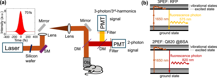
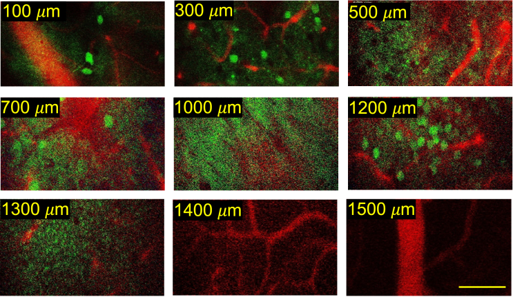
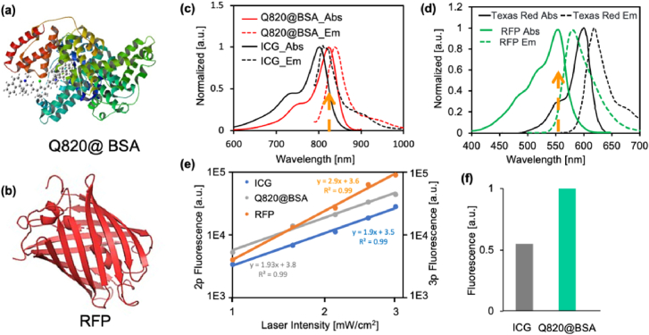
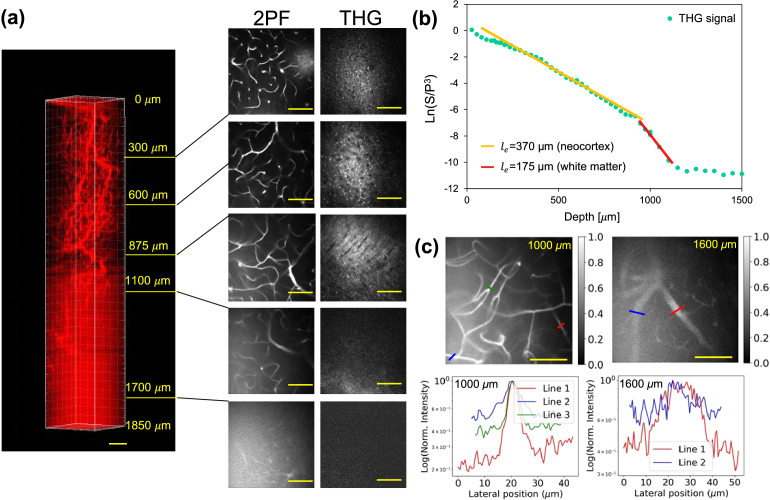
imaging of the neurovascular network is considered to be one of the most powerful approaches for understanding brain functionality. Nevertheless, simultaneously imaging the biological neural network and blood vessels in deep brain layers in a non-invasive manner remains to a major challenge due to the lack of appropriate labeling fluorescence probe pairs. Herein, we proposed a 2-photon and 3-photon fluorescence probe pair for neurovascular imaging. Specifically, the red fluorescence protein (RFP) with an absorption maximum of around 550 nm is used as a 3-photon excited probe to label neurons, and a cyanine derivative dye Q820@BSA has a NIR absorption maximum of 825 nm as a 2-photon excited probe to label the vasculature, enabling single wavelength excitation at 1650 nm for neurovascular imaging with high emission spectral separation (>250 nm). In particular, the 2-photon action cross-section of Q820@BSA was found to be about 2-fold larger than that of indocyanine green (ICG), a commonly used red 2-photon fluorescence labeling agent, at the same excitation wavelength. Benefiting from the long wavelength advantage in reducing scattering in both 2 and 3-photon excitation of the fluorescence pairs, we demonstrated neurovascular imaging in intact adult mouse brains through white matter and deep into the hippocampus in the somatosensory cortex.
神经血管网络成像被认为是理解大脑功能最有效的方法之一。然而,由于缺乏合适的标记荧光探针组,以非侵入性方式同时对大脑深层的生物神经网络和血管进行成像仍然是一个重大挑战。在此,我们提出了一种用于神经血管成像的双光子和三光子荧光探针组。具体而言,最大吸收波长约为550 nm的红色荧光蛋白(RFP)用作三光子激发探针来标记神经元,而一种花青衍生物染料Q820@BSA作为双光子激发探针,其近红外最大吸收波长为825 nm,用于标记脉管系统,能够在1650 nm进行单波长激发以实现具有高发射光谱分离(>250 nm)的神经血管成像。特别地,发现在相同激发波长下,Q820@BSA的双光子作用截面比常用的红色双光子荧光标记剂吲哚菁绿(ICG)大约2倍。受益于荧光对在双光子和三光子激发中减少散射的长波长优势,我们在完整的成年小鼠大脑中通过白质并深入体感皮层的海马体进行了神经血管成像。