• 文献检索
  • 文档翻译
  • 深度研究
  • 学术资讯
  • Suppr Zotero 插件Zotero 插件
  • 邀请有礼
  • 套餐&价格
  • 历史记录
应用&插件
Suppr Zotero 插件Zotero 插件浏览器插件Mac 客户端Windows 客户端微信小程序
定价
高级版会员购买积分包购买API积分包
服务
文献检索文档翻译深度研究API 文档MCP 服务
关于我们
关于 Suppr公司介绍联系我们用户协议隐私条款
关注我们

Suppr 超能文献

核心技术专利:CN118964589B侵权必究
粤ICP备2023148730 号-1Suppr @ 2026

文献检索

告别复杂PubMed语法,用中文像聊天一样搜索,搜遍4000万医学文献。AI智能推荐,让科研检索更轻松。

立即免费搜索

文件翻译

保留排版,准确专业,支持PDF/Word/PPT等文件格式,支持 12+语言互译。

免费翻译文档

深度研究

AI帮你快速写综述,25分钟生成高质量综述,智能提取关键信息,辅助科研写作。

立即免费体验

旁中心微视野视网膜敏感度作为与色素性视网膜炎视力丧失相关的关键参数

Parafoveal Microperimetric Retinal Sensitivity as a Key Parameter Associated with Vision Loss in Retinitis Pigmentosa.

作者信息

Hsiao Yu-Ting, Huang Hsiu-Mei, Chen Ta-Ching, Lo Jung, Chen Yung-Jen, Kuo Hsi-Kung, Lee Jong-Jer

机构信息

Department of Ophthalmology, Kaohsiung Chang Gung Memorial Hospital and Chang Gung University College of Medicine, Kaohsiung 83340, Taiwan.

Department of Ophthalmology, National Taiwan University Hospital, Taipei 10041, Taiwan.

出版信息

Diagnostics (Basel). 2024 Nov 29;14(23):2691. doi: 10.3390/diagnostics14232691.

DOI:10.3390/diagnostics14232691
PMID:39682599
原文链接:https://pmc.ncbi.nlm.nih.gov/articles/PMC11639803/
Abstract

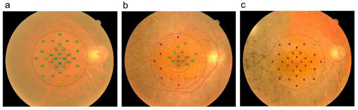
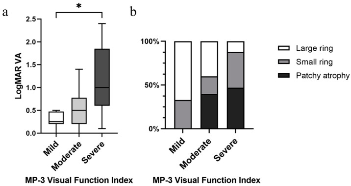
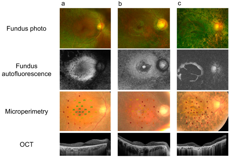
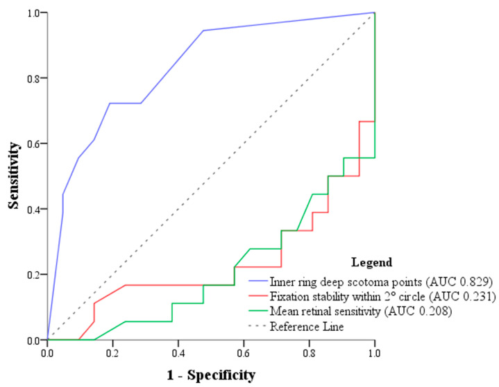
Although optical coherence tomography (OCT) is useful in determining outer retinal architecture, it may be suboptimal when monitoring subtle changes in retinitis pigmentosa (RP) patients. The aim of this study is to investigate precise microperimetric parameters for disease severity identification in RP patients. A cross-sectional and retrospective study. Thirty-nine eyes of 39 RP patients were included. Associations between logMAR visual acuity (VA), spectral-domain OCT, fundus autofluorescence imaging (FAF), and various microperimetric measures were evaluated. Microperimetric test locations were grouped into "foveal", parafoveal "inner ring", and perifoveal "outer ring". Independent variables were analyzed based on logistic regression, then assessed using area under the receiver operating characteristic curve (AUROC). Among all microperimetric measures, linear regression analysis indicated that mean retinal sensitivity and deep scotoma count at the parafoveal inner ring were the principal parameters associated with decreased VA. The AUROC was highest for deep scotoma count at the inner ring at a value of 0.829, with the cut-off point at 3.5. A visual function index was then established according to the number of parafoveal deep scotoma points, in order of mild (0 points), moderate (1-3 points), and severe (4 or more points). Our microperimetric visual function index also correlated significantly to logMAR VA and previously established FAF patterns. Our study discovered deep scotoma count at the parafoveal inner ring to be a key microperimetric parameter in evaluating vision loss in RP patients. Those with four or more deep scotoma points at the parafoveal inner ring are more likely to have functional low vision.

摘要

尽管光学相干断层扫描(OCT)有助于确定视网膜外层结构,但在监测视网膜色素变性(RP)患者的细微变化时可能并不理想。本研究的目的是探讨用于识别RP患者疾病严重程度的精确微视野参数。一项横断面回顾性研究。纳入了39例RP患者的39只眼。评估了对数最小分辨角视力(VA)、光谱域OCT、眼底自发荧光成像(FAF)与各种微视野测量之间的关联。微视野测试位置分为“中央凹”、中央凹旁“内环”和中央凹周围“外环”。基于逻辑回归分析自变量,然后使用受试者操作特征曲线下面积(AUROC)进行评估。在所有微视野测量中,线性回归分析表明,中央凹旁内环的平均视网膜敏感度和深部暗点计数是与视力下降相关的主要参数。内环深部暗点计数的AUROC最高,为0.829,截断点为3.5。然后根据中央凹旁深部暗点的数量建立了一个视觉功能指数,分为轻度(0个点)、中度(1 - 3个点)和重度(4个或更多点)。我们的微视野视觉功能指数也与对数最小分辨角视力和先前建立的FAF模式显著相关。我们的研究发现,中央凹旁内环的深部暗点计数是评估RP患者视力丧失的关键微视野参数。中央凹旁内环有四个或更多深部暗点的患者更有可能出现功能性低视力。

https://cdn.ncbi.nlm.nih.gov/pmc/blobs/1dcf/11639803/196a45d43af9/diagnostics-14-02691-g004.jpg
https://cdn.ncbi.nlm.nih.gov/pmc/blobs/1dcf/11639803/f0c9ba012a50/diagnostics-14-02691-g001.jpg
https://cdn.ncbi.nlm.nih.gov/pmc/blobs/1dcf/11639803/ba71c1fd5d15/diagnostics-14-02691-g002.jpg
https://cdn.ncbi.nlm.nih.gov/pmc/blobs/1dcf/11639803/eaabe5295eb8/diagnostics-14-02691-g003.jpg
https://cdn.ncbi.nlm.nih.gov/pmc/blobs/1dcf/11639803/196a45d43af9/diagnostics-14-02691-g004.jpg

相似文献

1
Parafoveal Microperimetric Retinal Sensitivity as a Key Parameter Associated with Vision Loss in Retinitis Pigmentosa.旁中心微视野视网膜敏感度作为与色素性视网膜炎视力丧失相关的关键参数
Diagnostics (Basel). 2024 Nov 29;14(23):2691. doi: 10.3390/diagnostics14232691.
2
Microperimetric correlations of autofluorescence and optical coherence tomography imaging in dry age-related macular degeneration.干性年龄相关性黄斑变性的自发荧光与光相干断层扫描成像的微视野相关性。
Am J Ophthalmol. 2012 Jun;153(6):1110-5. doi: 10.1016/j.ajo.2011.11.002. Epub 2012 Feb 8.
3
Multimodal Imaging in Retinitis Pigmentosa: Correlations among Microvascular Changes, Macular Function and Retinal Structure.视网膜色素变性的多模态成像:微血管变化、黄斑功能与视网膜结构之间的相关性
J Curr Ophthalmol. 2020 Apr 30;32(2):170-177. doi: 10.4103/JOCO.JOCO_99_20. eCollection 2020 Apr-Jun.
4
Evaluation of Structural Retinal Layer Alterations in Retinitis Pigmentosa.评估视网膜色素变性的结构视网膜层改变。
Rom J Ophthalmol. 2023 Oct-Dec;67(4):326-336. doi: 10.22336/rjo.2023.53.
5
Diagnostic imaging in patients with retinitis pigmentosa.视网膜色素变性患者的诊断性影像学检查
J Med Invest. 2012;59(1-2):1-11. doi: 10.2152/jmi.59.1.
6
Correlation between macular morphology and sensitivity in patients with retinitis pigmentosa and hyperautofluorescent ring.色素性视网膜炎和高自发荧光环患者的黄斑形态与敏感性的相关性。
Invest Ophthalmol Vis Sci. 2012 Jan 3;53(1):47-52. doi: 10.1167/iovs.11-8048.
7
Changes of fundus autofluorescence, photoreceptor inner and outer segment junction line, and visual function in patients with retinitis pigmentosa.色素性视网膜炎患者的眼底自发荧光、光感受器内外节连接带和视觉功能的变化。
Clin Exp Ophthalmol. 2010 Aug;38(6):597-604. doi: 10.1111/j.1442-9071.2010.02321.x. Epub 2010 Apr 29.
8
Quantification of Macular Microvascular Changes in Retinitis Pigmentosa Using Optical Coherence Tomography Angiography.使用光学相干断层扫描血管造影术对视网膜色素变性黄斑部微血管变化进行定量分析。
Clin Ophthalmol. 2020 Jun 22;14:1705-1713. doi: 10.2147/OPTH.S254909. eCollection 2020.
9
Autofluorescence imaging and spectral-domain optical coherence tomography in incomplete congenital stationary night blindness and comparison with retinitis pigmentosa.先天性静止性夜盲不完全型的自发荧光成像和谱域光相干断层扫描,并与视网膜色素变性进行比较。
Am J Ophthalmol. 2012 Jan;153(1):143-54.e2. doi: 10.1016/j.ajo.2011.06.018. Epub 2011 Sep 13.
10
Fundus autofluorescence and retinal structure as determined by spectral domain optical coherence tomography, and retinal function in retinitis pigmentosa.眼底自发荧光和光谱域光相干断层扫描所确定的视网膜结构,以及色素性视网膜炎的视网膜功能。
Graefes Arch Clin Exp Ophthalmol. 2012 Mar;250(3):333-9. doi: 10.1007/s00417-011-1823-5. Epub 2011 Sep 24.

本文引用的文献

1
Multimodal Imaging of Optic Nerve Head in Retinitis Pigmentosa.色素性视网膜炎的视神经头的多模态成像。
Semin Ophthalmol. 2024 Aug;39(6):480-487. doi: 10.1080/08820538.2024.2363849. Epub 2024 Jun 8.
2
In-Depth Retinal Sensitivity Assessment With the MP3 Type S Microperimeter: A Methods Study.用 MP3 型 S 微视野计进行深入的视网膜灵敏度评估:一种方法研究。
Transl Vis Sci Technol. 2024 Apr 2;13(4):14. doi: 10.1167/tvst.13.4.14.
3
Comparison of Novel Volumetric Microperimetry Metrics in Intermediate Age-Related Macular Degeneration: PINNACLE Study Report 3.
新型中龄相关性黄斑变性容积微视野计测量指标的比较:PINNACLE 研究报告 3。
Transl Vis Sci Technol. 2023 Aug 1;12(8):21. doi: 10.1167/tvst.12.8.21.
4
Microperimetry as an Outcome Measure in RPGR-associated Retinitis Pigmentosa Clinical Trials.微视野计作为 RPGR 相关性视锥细胞营养不良临床试验的一种结局测量方法。
Transl Vis Sci Technol. 2023 Jun 1;12(6):4. doi: 10.1167/tvst.12.6.4.
5
A cross-sectional study to assess the clinical utility of modern visual function assessments in patients with inherited retinal disease: a mixed methods observational study protocol.一项横断面研究,旨在评估现代视觉功能评估在遗传性视网膜疾病患者中的临床应用价值:一项混合方法观察研究方案。
BMC Ophthalmol. 2023 May 24;23(1):234. doi: 10.1186/s12886-023-02974-6.
6
Microperimetry in Retinal Diseases.眼底病的微视野检查
Asia Pac J Ophthalmol (Phila). 2023;12(2):211-227. doi: 10.1097/APO.0000000000000597. Epub 2023 Mar 24.
7
Baseline Microperimetry and OCT in the RUSH2A Study: Structure-Function Association and Correlation With Disease Severity.RUSH2A 研究中的基线微视野计和 OCT:结构-功能相关性及其与疾病严重程度的关系。
Am J Ophthalmol. 2022 Dec;244:98-116. doi: 10.1016/j.ajo.2022.08.013. Epub 2022 Aug 22.
8
Characterizing Visual Fields in RPGR Related Retinitis Pigmentosa Using Octopus Static-Automated Perimetry.使用欧宝静态自动化视野计描述 RPGR 相关性视锥细胞营养不良的视野特征。
Transl Vis Sci Technol. 2022 May 2;11(5):15. doi: 10.1167/tvst.11.5.15.
9
Genetic testing and diagnosis of inherited retinal diseases.遗传性视网膜疾病的基因检测和诊断。
Orphanet J Rare Dis. 2021 Dec 14;16(1):514. doi: 10.1186/s13023-021-02145-0.
10
The structure-function correlation analysed by OCT and full field ERG in typical and pericentral subtypes of retinitis pigmentosa.经 OCT 和全视野 ERG 分析典型和旁中心型色素性视网膜炎的结构-功能相关性。
Sci Rep. 2021 Aug 19;11(1):16883. doi: 10.1038/s41598-021-96570-7.