Zhao Xiaojiang, Wang Yuhang, Xue Mengli, Ding Yun, Zhang Han, Wang Kai, Ren Jie, Li Xin, Xu Meilin, Lv Jun, Wang Zixiao, Sun Daqiang
Chest hospital, Tianjin University, Tianjin, China.
Department of Thoracic Surgery, Tianjin Chest Hospital, No. 261, Taierzhuang South Road, Jinnan District, Tianjin, 300222, China.
Cancer Imaging. 2024 Dec 18;24(1):167. doi: 10.1186/s40644-024-00813-5.
To develop a multimodal predictive model, Radiomics Integrated TLSs System (RAITS), based on preoperative CT radiomic features for the identification of TLSs in stage I lung adenocarcinoma patients and to evaluate its potential in prognosis stratification and guiding personalized treatment.
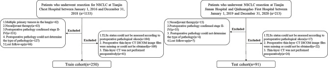
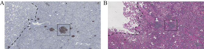
The most recent preoperative chest CT thin-slice scans and postoperative hematoxylin and eosin-stained pathology sections of patients diagnosed with stage I LUAD were retrospectively collected. Tumor segmentation was achieved using an automatic virtual adversarial training segmentation algorithm based on a three-dimensional U-shape convolutional neural network (3D U-Net). Radiomic features were extracted from the tumor and peritumoral areas, with extensions of 2 mm, 4 mm, 6 mm, and 8 mm, respectively, and deep learning image features were extracted through a convolutional neural network. Subsequently, the RAITS was constructed. The performance of RAITS was then evaluated in both the train and validation cohorts.
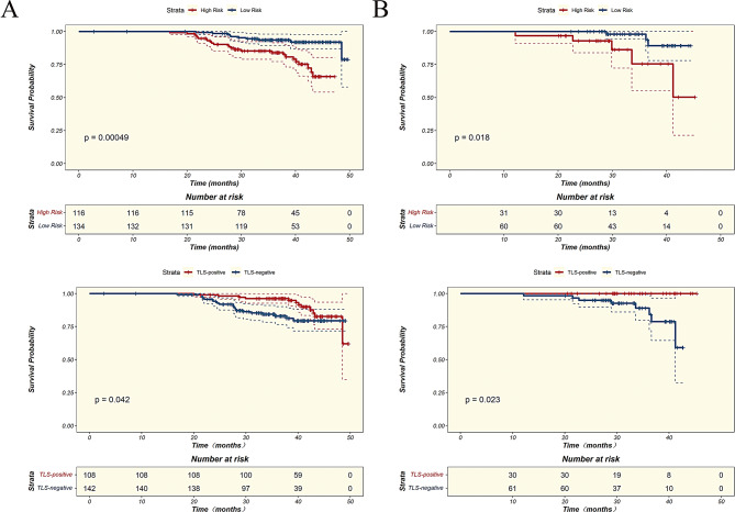
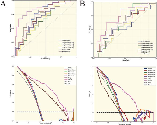
RAITS demonstrated superior AUC, sensitivity, and specificity in both the training and external validation cohorts, outperforming traditional unimodal models. In the validation cohort, RAITS achieved an AUC of 0.78 (95% CI, 0.69-0.88) and showed higher net benefits across most threshold ranges. RAITS exhibited strong discriminative ability in risk stratification, with p < 0.01 in the training cohort and p = 0.02 in the validation cohort, consistent with the actual predictive performance of TLSs, where TLS-positive patients had significantly higher recurrence-free survival (RFS) compared to TLS-negative patients (p = 0.04 in the training cohort, p = 0.02 in the validation cohort).
As a multimodal predictive model based on preoperative CT radiomic features, RAITS demonstrated excellent performance in identifying TLSs in stage I LUAD and holds potential value in clinical decision-making.
基于术前CT影像组学特征开发一种多模态预测模型——影像组学整合三级淋巴结构系统(RAITS),用于识别I期肺腺癌患者的三级淋巴结构(TLSs),并评估其在预后分层和指导个性化治疗方面的潜力。
回顾性收集诊断为I期肺腺癌患者的最新术前胸部CT薄层扫描及术后苏木精-伊红染色病理切片。使用基于三维U型卷积神经网络(3D U-Net)的自动虚拟对抗训练分割算法进行肿瘤分割。从肿瘤及瘤周区域分别提取扩展2毫米、4毫米、6毫米和8毫米的影像组学特征,并通过卷积神经网络提取深度学习图像特征。随后构建RAITS。然后在训练队列和验证队列中评估RAITS的性能。
RAITS在训练队列和外部验证队列中均表现出优异的曲线下面积(AUC)、敏感性和特异性,优于传统单模态模型。在验证队列中,RAITS的AUC为0.78(95%置信区间,0.69 - 0.88),并且在大多数阈值范围内显示出更高的净效益。RAITS在风险分层中表现出很强的判别能力,训练队列中p < 0.01,验证队列中p = 0.02,与TLSs的实际预测性能一致,其中TLS阳性患者的无复发生存期(RFS)显著高于TLS阴性患者(训练队列中p = 0.04,验证队列中p = 0.02)。
作为一种基于术前CT影像组学特征的多模态预测模型,RAITS在识别I期肺腺癌中的TLSs方面表现出优异的性能,在临床决策中具有潜在价值。