• 文献检索
  • 文档翻译
  • 深度研究
  • 学术资讯
  • Suppr Zotero 插件Zotero 插件
  • 邀请有礼
  • 套餐&价格
  • 历史记录
应用&插件
Suppr Zotero 插件Zotero 插件浏览器插件Mac 客户端Windows 客户端微信小程序
定价
高级版会员购买积分包购买API积分包
服务
文献检索文档翻译深度研究API 文档MCP 服务
关于我们
关于 Suppr公司介绍联系我们用户协议隐私条款
关注我们

Suppr 超能文献

核心技术专利:CN118964589B侵权必究
粤ICP备2023148730 号-1Suppr @ 2026

文献检索

告别复杂PubMed语法,用中文像聊天一样搜索,搜遍4000万医学文献。AI智能推荐,让科研检索更轻松。

立即免费搜索

文件翻译

保留排版,准确专业,支持PDF/Word/PPT等文件格式,支持 12+语言互译。

免费翻译文档

深度研究

AI帮你快速写综述,25分钟生成高质量综述,智能提取关键信息,辅助科研写作。

立即免费体验

一种多空间信息表征模型,强调利用结构磁共振成像进行阿尔茨海默病诊断的关键脑区。

A multispatial information representation model emphasizing key brain regions for Alzheimer's disease diagnosis with structural magnetic resonance imaging.

作者信息

Nan Pengzhi, Li Lin, Song Zhiwei, Wang Yi, Zhu Chuanzhen, Hu Fang, Zheng Qiang

机构信息

School of Computer and Control Engineering, Yantai University, Yantai, China.

Department of Radiology, Yantaishan Hospital Affiliated to Binzhou Medical University, Yantai, China.

出版信息

Quant Imaging Med Surg. 2024 Dec 5;14(12):8568-8585. doi: 10.21037/qims-24-584. Epub 2024 Nov 29.

DOI:10.21037/qims-24-584
PMID:39698706
原文链接:https://pmc.ncbi.nlm.nih.gov/articles/PMC11651950/
Abstract

BACKGROUND

Structural magnetic resonance imaging (sMRI) can reflect structural abnormalities of the brain. Due to its high tissue contrast and spatial resolution, it is considered as an MRI sequence in diagnostic tasks related to Alzheimer's disease (AD). Thus far, most studies based on sMRI have only focused on pathological changes in disease-related brain regions in Euclidean space, ignoring the association and interaction between brain regions represented in non-Euclidean space. This non-Euclidean spatial information can provide valuable information for brain disease research. However, few studies have combined Euclidean spatial information in images and graph spatial information in brain networks for the early diagnosis of AD. The purpose of this study is to explore how to effectively combine multispatial information for enhancing AD diagnostic performance.

METHODS

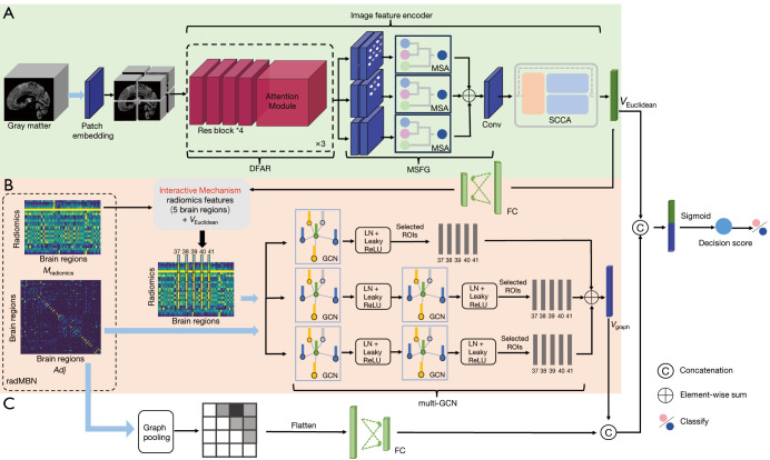
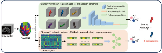
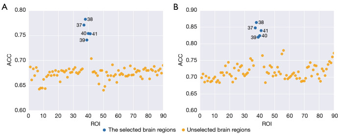
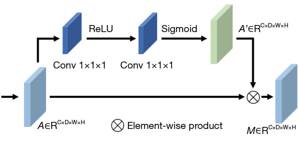
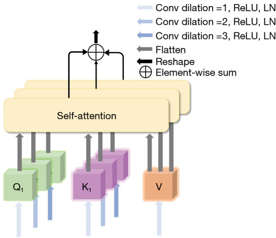
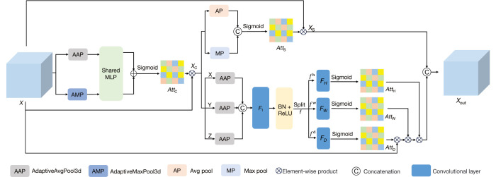
A multispatial information representation model (MSRNet) was constructed for the diagnosis of AD using sMRI. Specifically, the MSRNet included a Euclidean representation channel integrating a multiscale module and a feature enhancement module, in addition to a graph (non-Euclidean) representation channel integrating a node feature aggregation mechanism. This was accomplished through the adoption of a multilayer graph convolutional neural network and a node connectivity aggregation mechanism with fully connected layers. Each participants' gray-matter volume map and preconstructed radiomics-based morphology brain network (radMBN) were used as MSRNet inputs for the learning of multispatial information. Other than the multispatial information representation in MSRNet, an interactive mechanism was proposed to connect the Euclidean and graph representation channels by five disease-related brain regions which were identified based on a classifier operated on with two feature strategies of voxel intensities and radiomics features. MSRNet focused on disease-related brain regions while integrating multispatial information to effectively enhance disease discrimination.

RESULTS

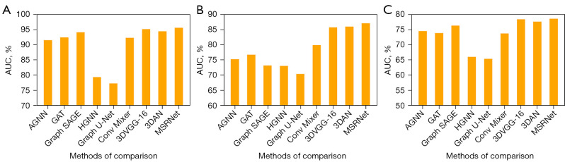
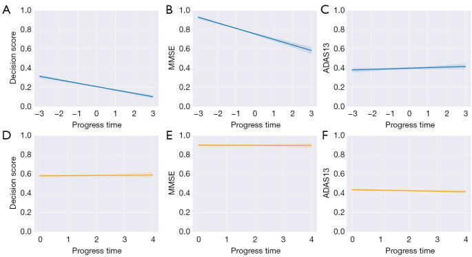
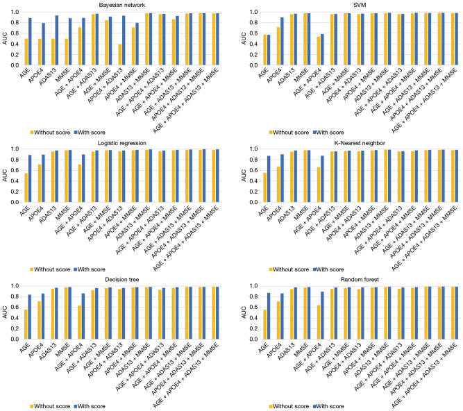
The MSRNet was validated on four publicly available datasets, achieving accuracies 92.8% and 90.6% for AD in intra-database and inter-database cross-validation, respectively. The accuracy of MSRNet in distinguishing between late mild cognitive impairment (MCI) and early MCI, and between progressive MCI and stable MCI, reached 79.8% and 73.4%, respectively. The experiments demonstrated that the model's decision scores exhibited good detection capability for MCI progression. Furthermore, the potential of decision scores for improving diagnostic performance was exhibited by combining decision scores with other clinical indicators for AD identification.

CONCLUSIONS

The MSRNet model could conduct an effective multispatial information representation in the sMRI-based diagnosis of AD. The proposed interaction mechanism in the MSRNet could help the model focus on AD-related brain regions, thus further improving the diagnostic ability.

摘要

背景

结构磁共振成像(sMRI)能够反映大脑的结构异常。由于其具有高组织对比度和空间分辨率,它被视为用于与阿尔茨海默病(AD)相关诊断任务的MRI序列。到目前为止,大多数基于sMRI的研究仅关注欧几里得空间中疾病相关脑区的病理变化,而忽略了非欧几里得空间中所代表的脑区之间的关联和相互作用。这种非欧几里得空间信息可为脑部疾病研究提供有价值的信息。然而,很少有研究将图像中的欧几里得空间信息和脑网络中的图空间信息结合起来用于AD的早期诊断。本研究的目的是探索如何有效地结合多空间信息以提高AD诊断性能。

方法

构建了一个用于利用sMRI诊断AD的多空间信息表示模型(MSRNet)。具体而言,MSRNet除了包括一个整合节点特征聚合机制的图(非欧几里得)表示通道外,还包括一个整合多尺度模块和特征增强模块的欧几里得表示通道。这是通过采用多层图卷积神经网络和具有全连接层的节点连通性聚合机制来实现的。将每个参与者的灰质体积图和预先构建的基于放射组学的形态学脑网络(radMBN)用作MSRNet的输入,以学习多空间信息。除了MSRNet中的多空间信息表示外,还提出了一种交互机制,通过基于体素强度和放射组学特征的两种特征策略操作的分类器所确定的五个与疾病相关的脑区来连接欧几里得和图表示通道。MSRNet在整合多空间信息的同时关注与疾病相关的脑区,以有效增强疾病区分能力。

结果

MSRNet在四个公开可用数据集上得到验证,在数据库内和数据库间交叉验证中,AD的准确率分别达到92.8%和90.6%。MSRNet区分晚期轻度认知障碍(MCI)和早期MCI以及区分进展性MCI和稳定MCI的准确率分别达到79.8%和73.4%。实验表明,该模型的决策分数对MCI进展具有良好的检测能力。此外,通过将决策分数与用于AD识别的其他临床指标相结合,展示了决策分数在提高诊断性能方面的潜力。

结论

MSRNet模型在基于sMRI的AD诊断中能够进行有效的多空间信息表示。MSRNet中提出的交互机制可帮助模型关注与AD相关的脑区,从而进一步提高诊断能力。

https://cdn.ncbi.nlm.nih.gov/pmc/blobs/0c3d/11651950/14838585ca55/qims-14-12-8568-f10.jpg
https://cdn.ncbi.nlm.nih.gov/pmc/blobs/0c3d/11651950/6f583cb69ffd/qims-14-12-8568-f1.jpg
https://cdn.ncbi.nlm.nih.gov/pmc/blobs/0c3d/11651950/cca0de99faec/qims-14-12-8568-f2.jpg
https://cdn.ncbi.nlm.nih.gov/pmc/blobs/0c3d/11651950/b7f12e865688/qims-14-12-8568-f3.jpg
https://cdn.ncbi.nlm.nih.gov/pmc/blobs/0c3d/11651950/f9d4c3d3a77f/qims-14-12-8568-f4.jpg
https://cdn.ncbi.nlm.nih.gov/pmc/blobs/0c3d/11651950/755be10b1954/qims-14-12-8568-f5.jpg
https://cdn.ncbi.nlm.nih.gov/pmc/blobs/0c3d/11651950/df865eddc8c5/qims-14-12-8568-f6.jpg
https://cdn.ncbi.nlm.nih.gov/pmc/blobs/0c3d/11651950/137362d15497/qims-14-12-8568-f7.jpg
https://cdn.ncbi.nlm.nih.gov/pmc/blobs/0c3d/11651950/a022b3164db8/qims-14-12-8568-f8.jpg
https://cdn.ncbi.nlm.nih.gov/pmc/blobs/0c3d/11651950/beef251ce452/qims-14-12-8568-f9.jpg
https://cdn.ncbi.nlm.nih.gov/pmc/blobs/0c3d/11651950/14838585ca55/qims-14-12-8568-f10.jpg

相似文献

1
A multispatial information representation model emphasizing key brain regions for Alzheimer's disease diagnosis with structural magnetic resonance imaging.一种多空间信息表征模型,强调利用结构磁共振成像进行阿尔茨海默病诊断的关键脑区。
Quant Imaging Med Surg. 2024 Dec 5;14(12):8568-8585. doi: 10.21037/qims-24-584. Epub 2024 Nov 29.
2
sMRI-ADNet: an interpretable deep learning framework integrating Euclidean-graph representations of Alzheimer's disease solely from structural MRI.sMRI-ADNet:一种可解释的深度学习框架,仅从结构 MRI 中整合阿尔茨海默病的欧几里得图表示。
MAGMA. 2024 Oct;37(5):845-857. doi: 10.1007/s10334-024-01178-3. Epub 2024 Jun 13.
3
Jointly constrained group sparse connectivity representation improves early diagnosis of Alzheimer's disease on routinely acquired T1-weighted imaging-based brain network.联合约束组稀疏连通性表示法可改善基于常规获取的T1加权成像脑网络的阿尔茨海默病早期诊断。
Health Inf Sci Syst. 2024 Mar 6;12(1):19. doi: 10.1007/s13755-023-00269-0. eCollection 2024 Dec.
4
Alzheimer's Disease Diagnosis and Biomarker Analysis Using Resting-State Functional MRI Functional Brain Network With Multi-Measures Features and Hippocampal Subfield and Amygdala Volume of Structural MRI.使用具有多测量特征的静息态功能磁共振成像功能脑网络以及结构磁共振成像的海马亚区和杏仁核体积进行阿尔茨海默病诊断和生物标志物分析
Front Aging Neurosci. 2022 May 30;14:818871. doi: 10.3389/fnagi.2022.818871. eCollection 2022.
5
Graph Convolutional Network for AD and MCI Diagnosis Utilizing Peripheral DNA Methylation: Réseau de neurones en graphes pour le diagnostic de la MA et du TCL à l'aide de la méthylation de l'ADN périphérique.利用外周血DNA甲基化的阿尔茨海默病和轻度认知障碍诊断的图卷积网络:使用外周血DNA甲基化进行阿尔茨海默病和轻度认知障碍诊断的图神经网络
Can J Psychiatry. 2024 Dec;69(12):869-879. doi: 10.1177/07067437241300947. Epub 2024 Nov 25.
6
THAN: task-driven hierarchical attention network for the diagnosis of mild cognitive impairment and Alzheimer's disease.THAN:用于轻度认知障碍和阿尔茨海默病诊断的任务驱动分层注意力网络
Quant Imaging Med Surg. 2021 Jul;11(7):3338-3354. doi: 10.21037/qims-21-91.
7
Early Detection of Alzheimer's Disease Using Magnetic Resonance Imaging: A Novel Approach Combining Convolutional Neural Networks and Ensemble Learning.利用磁共振成像早期检测阿尔茨海默病:一种结合卷积神经网络和集成学习的新方法
Front Neurosci. 2020 May 13;14:259. doi: 10.3389/fnins.2020.00259. eCollection 2020.
8
Multimodal feature fusion-based graph convolutional networks for Alzheimer's disease stage classification using F-18 florbetaben brain PET images and clinical indicators.基于多模态特征融合的图卷积网络用于使用F-18氟贝他班脑PET图像和临床指标的阿尔茨海默病阶段分类
PLoS One. 2024 Dec 23;19(12):e0315809. doi: 10.1371/journal.pone.0315809. eCollection 2024.
9
An extensible hierarchical graph convolutional network for early Alzheimer's disease identification.用于早期阿尔茨海默病识别的可扩展分层图卷积网络。
Comput Methods Programs Biomed. 2023 Aug;238:107597. doi: 10.1016/j.cmpb.2023.107597. Epub 2023 May 15.
10
Predicting MCI to AD Conversation Using Integrated sMRI and rs-fMRI: Machine Learning and Graph Theory Approach.使用整合的结构磁共振成像和静息态功能磁共振成像预测轻度认知障碍向阿尔茨海默病的转变:机器学习与图论方法
Front Aging Neurosci. 2021 Jul 30;13:688926. doi: 10.3389/fnagi.2021.688926. eCollection 2021.

本文引用的文献

1
CAT: a computational anatomy toolbox for the analysis of structural MRI data.CAT:用于分析结构磁共振成像数据的计算解剖工具箱。
Gigascience. 2024 Jan 2;13. doi: 10.1093/gigascience/giae049.
2
Morphological connectivity differences in Alzheimer's disease correlate with gene transcription and cell-type.阿尔茨海默病患者的形态连通性差异与基因转录和细胞类型相关。
Hum Brain Mapp. 2023 Dec 15;44(18):6364-6374. doi: 10.1002/hbm.26512. Epub 2023 Oct 17.
3
Deep joint learning of pathological region localization and Alzheimer's disease diagnosis.
病理性区域定位与阿尔茨海默病诊断的深度联合学习。
Sci Rep. 2023 Jul 19;13(1):11664. doi: 10.1038/s41598-023-38240-4.
4
Multi-modal cross-attention network for Alzheimer's disease diagnosis with multi-modality data.多模态跨注意网络用于基于多模态数据的阿尔茨海默病诊断。
Comput Biol Med. 2023 Aug;162:107050. doi: 10.1016/j.compbiomed.2023.107050. Epub 2023 May 22.
5
An extensible hierarchical graph convolutional network for early Alzheimer's disease identification.用于早期阿尔茨海默病识别的可扩展分层图卷积网络。
Comput Methods Programs Biomed. 2023 Aug;238:107597. doi: 10.1016/j.cmpb.2023.107597. Epub 2023 May 15.
6
A comprehensive characterization of hippocampal feature ensemble serves as individualized brain signature for Alzheimer's disease: deep learning analysis in 3238 participants worldwide.海马体特征集合的全面表征作为阿尔茨海默病的个体化脑特征:全球3238名参与者的深度学习分析
Eur Radiol. 2023 Aug;33(8):5385-5397. doi: 10.1007/s00330-023-09519-x. Epub 2023 Mar 9.
7
VGG-TSwinformer: Transformer-based deep learning model for early Alzheimer's disease prediction.VGG-TSwinformer:基于 Transformer 的深度学习模型,用于早期阿尔茨海默病预测。
Comput Methods Programs Biomed. 2023 Feb;229:107291. doi: 10.1016/j.cmpb.2022.107291. Epub 2022 Nov 30.
8
Regional Radiomics Similarity Networks Reveal Distinct Subtypes and Abnormality Patterns in Mild Cognitive Impairment.区域放射组学相似性网络揭示轻度认知障碍的不同亚型和异常模式。
Adv Sci (Weinh). 2022 Apr;9(12):e2104538. doi: 10.1002/advs.202104538. Epub 2022 Jan 31.
9
Regional radiomics similarity networks (R2SNs) in the human brain: Reproducibility, small-world properties and a biological basis.人类大脑中的区域放射组学相似性网络(R2SNs):可重复性、小世界特性及生物学基础。
Netw Neurosci. 2021 Aug 30;5(3):783-797. doi: 10.1162/netn_a_00200. eCollection 2021.
10
Screening and predicting progression from high-risk mild cognitive impairment to Alzheimer's disease.筛查和预测高危轻度认知障碍向阿尔茨海默病的进展。
Sci Rep. 2021 Sep 2;11(1):17558. doi: 10.1038/s41598-021-96914-3.