• 文献检索
  • 文档翻译
  • 深度研究
  • 学术资讯
  • Suppr Zotero 插件Zotero 插件
  • 邀请有礼
  • 套餐&价格
  • 历史记录
应用&插件
Suppr Zotero 插件Zotero 插件浏览器插件Mac 客户端Windows 客户端微信小程序
定价
高级版会员购买积分包购买API积分包
服务
文献检索文档翻译深度研究API 文档MCP 服务
关于我们
关于 Suppr公司介绍联系我们用户协议隐私条款
关注我们

Suppr 超能文献

核心技术专利:CN118964589B侵权必究
粤ICP备2023148730 号-1Suppr @ 2026

文献检索

告别复杂PubMed语法,用中文像聊天一样搜索,搜遍4000万医学文献。AI智能推荐,让科研检索更轻松。

立即免费搜索

文件翻译

保留排版,准确专业,支持PDF/Word/PPT等文件格式,支持 12+语言互译。

免费翻译文档

深度研究

AI帮你快速写综述,25分钟生成高质量综述,智能提取关键信息,辅助科研写作。

立即免费体验

动态对比增强磁共振成像在评估前列腺腺癌中的作用:一项对前列腺切除患者队列进行的部分盲法回顾性研究,该研究将全腺体组织病理学相关性与PI-RADS或TNM分期的应用相结合。

The Role of Dynamic Contrast Enhanced Magnetic Resonance Imaging in Evaluating Prostate Adenocarcinoma: A Partially-Blinded Retrospective Study of a Prostatectomy Patient Cohort With Whole Gland Histopathology Correlation and Application of PI-RADS or TNM Staging.

作者信息

Sridhar Sajeev, Abouelfetouh Zeyad, Codreanu Ion, Gupta Nakul, Zhang Shu, Efstathiou Eleni, Karolyi Daniel K, Shen Steven S, LaViolette Peter S, Miles Brian, Martin Diego R

机构信息

Department of Radiology, Houston Methodist Research Institute, Houston, Texas, USA.

Department of Radiology, Houston Methodist Research Institute, Nicolae Testemițanu State University of Medicine and Pharmacy, Chișinău, Moldova.

出版信息

Prostate. 2025 Apr;85(5):413-423. doi: 10.1002/pros.24843. Epub 2024 Dec 19.

DOI:10.1002/pros.24843
PMID:39702937
原文链接:https://pmc.ncbi.nlm.nih.gov/articles/PMC11848987/
Abstract

BACKGROUND

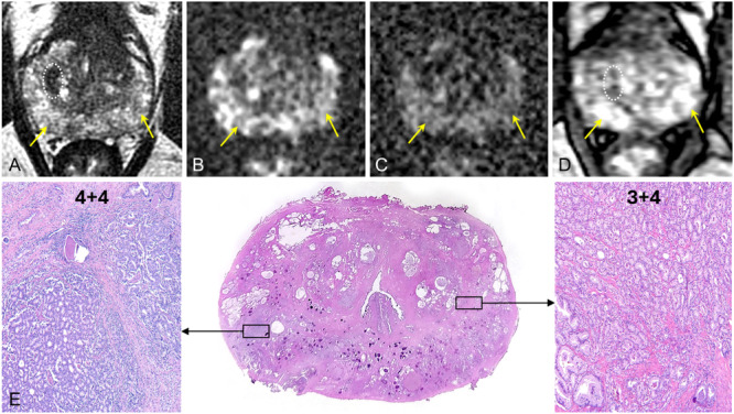
Dynamic contrast-enhanced (DCE) magnetic resonance imaging (MRI) in the current Prostate Imaging-Reporting and Data System version 2.1 (PI-RADS v2.1) is considered optional, with primary scoring based on T2-weighted imaging (T2WI) and diffusion weighted imaging (DWI). Our study is designed to assess the relative contribution of DCE MRI in a patient-cohort with whole mount prostate histopathology and spatially-mapped prostate adenocarcinoma (PCa) for reference.

METHODS

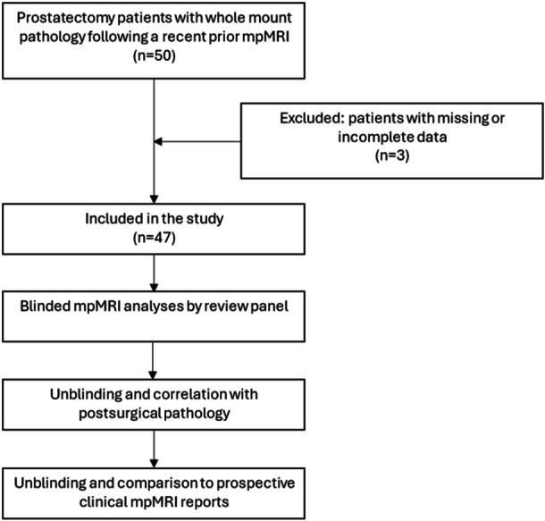
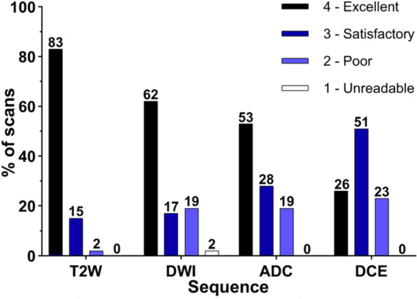
We performed a partially-blinded retrospective review of 47 prostatectomy patients with recent multi-parametric MRI (mpMRI). Scans included T2WI, DWI with apparent diffusion coefficient (ADC) mapping, and DCE imaging. Lesion conspicuity was scored on a 10-point scale with ≥ 6 considered "positive," and image quality was assessed on a 4-point scale for each sequence. The diagnostic contribution of DCE images was evaluated on a 4-point scale. The mpMRI studies were assigned PI-RADS scores and tumor, node, metastasis (TNM) T-stage with blinded comparison to spatially-mapped whole-mount pathology. Results were compared to the prospective clinical reports, which used standardized PI-RADS templates that emphasize T2WI, DWI and ADC.

RESULTS

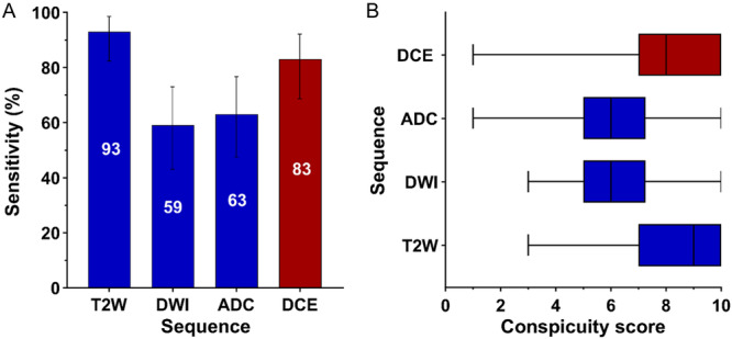
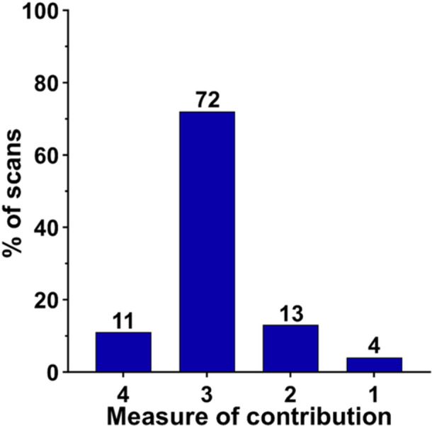
Per lesion sensitivity for PCa was 93.5%, 82.6%, 63.0%, and 58.7% on T2WI, DCE, ADC and DWI, respectively. Mean lesion conspicuity was 8.5, 7.9, 6.2, and 6.1, on T2W, DCE, ADC and DWI, respectively. The higher values on T2WI and DCE imaging were not significantly different from each other but were both significantly different from DWI and ADC (p < 0.001). DCE scans were determined to have a marked diagnostic contribution in 83% of patients, with the most common diagnostic yield being detection of contralateral peripheral zone tumor or delineating presence/absence of extra-prostatic extension (EPE), contributing to more accurate PCa staging by PI-RADS or TNM, as compared to histopathology.

CONCLUSION

We demonstrate that DCE may contribute to lesion detection and local staging as compared to T2WI plus DWI-ADC alone and that lesion conspicuity using DCE is markedly improved as compared to DWI-ADC. These findings support modification of PI-RADS v2.1 to include use of DCE acquisitions and that a TNM staging is feasible on mpMRI as compared to surgical pathology.

摘要

背景

在当前的前列腺影像报告和数据系统第2.1版(PI-RADS v2.1)中,动态对比增强(DCE)磁共振成像(MRI)被认为是可选的,主要评分基于T2加权成像(T2WI)和扩散加权成像(DWI)。我们的研究旨在评估DCE MRI在一组具有全层前列腺组织病理学和空间映射前列腺腺癌(PCa)的患者队列中的相对贡献以供参考。

方法

我们对47例近期接受多参数MRI(mpMRI)检查的前列腺切除患者进行了部分盲法回顾性研究。扫描包括T2WI、具有表观扩散系数(ADC)图的DWI和DCE成像。病变的明显程度采用10分制评分,≥6分被认为“阳性”,每个序列的图像质量采用4分制评估。DCE图像的诊断贡献采用4分制评估。mpMRI研究被赋予PI-RADS评分以及肿瘤、淋巴结、转移(TNM)T分期,并与空间映射的全层病理进行盲法比较。结果与前瞻性临床报告进行比较,前瞻性临床报告使用强调T2WI、DWI和ADC的标准化PI-RADS模板。

结果

PCa的每个病变敏感性在T2WI、DCE、ADC和DWI上分别为93.5%、82.6%、63.0%和58.7%。T2WI、DCE、ADC和DWI上病变的平均明显程度分别为8.5、7.9、6.2和6.1。T2WI和DCE成像上的较高值彼此之间无显著差异,但均与DWI和ADC有显著差异(p<0.001)。确定83%的患者DCE扫描具有显著的诊断贡献,最常见的诊断结果是检测对侧外周带肿瘤或确定是否存在前列腺外侵犯(EPE),与组织病理学相比,有助于通过PI-RADS或TNM更准确地对PCa进行分期。

结论

我们证明,与单独的T2WI加DWI-ADC相比,DCE可能有助于病变检测和局部分期,并且与DWI-ADC相比,使用DCE时病变的明显程度有显著改善。这些发现支持对PI-RADS v2.1进行修改以纳入DCE采集的使用,并且与手术病理相比,在mpMRI上进行TNM分期是可行的。

https://cdn.ncbi.nlm.nih.gov/pmc/blobs/8c4e/11848987/ca38d7cba585/PROS-85-413-g003.jpg
https://cdn.ncbi.nlm.nih.gov/pmc/blobs/8c4e/11848987/53c3ebaee5d5/PROS-85-413-g005.jpg
https://cdn.ncbi.nlm.nih.gov/pmc/blobs/8c4e/11848987/ddec7cece367/PROS-85-413-g002.jpg
https://cdn.ncbi.nlm.nih.gov/pmc/blobs/8c4e/11848987/3b8536dba6dc/PROS-85-413-g004.jpg
https://cdn.ncbi.nlm.nih.gov/pmc/blobs/8c4e/11848987/f50b1c051c50/PROS-85-413-g001.jpg
https://cdn.ncbi.nlm.nih.gov/pmc/blobs/8c4e/11848987/ca38d7cba585/PROS-85-413-g003.jpg

相似文献

1
The Role of Dynamic Contrast Enhanced Magnetic Resonance Imaging in Evaluating Prostate Adenocarcinoma: A Partially-Blinded Retrospective Study of a Prostatectomy Patient Cohort With Whole Gland Histopathology Correlation and Application of PI-RADS or TNM Staging.动态对比增强磁共振成像在评估前列腺腺癌中的作用:一项对前列腺切除患者队列进行的部分盲法回顾性研究,该研究将全腺体组织病理学相关性与PI-RADS或TNM分期的应用相结合。
Prostate. 2025 Apr;85(5):413-423. doi: 10.1002/pros.24843. Epub 2024 Dec 19.
2
Prospective evaluation of mpMRI-derived nomograms for detecting prostate cancer in PI-RADS v2.1 upgraded and non-upgraded lesions.基于多参数磁共振成像(mpMRI)的列线图对PI-RADS v2.1升级和未升级病变中前列腺癌检测的前瞻性评估
Front Oncol. 2025 Jun 4;15:1510049. doi: 10.3389/fonc.2025.1510049. eCollection 2025.
3
Preoperative Diagnosis of Ipsilateral and Contralateral Breast Cancer: Role of Diffusion-weighted MRI.同侧及对侧乳腺癌的术前诊断:扩散加权磁共振成像的作用
Radiology. 2025 Jun;315(3):e242423. doi: 10.1148/radiol.242423.
4
Magnetic resonance perfusion for differentiating low-grade from high-grade gliomas at first presentation.首次就诊时磁共振灌注成像用于鉴别低级别与高级别胶质瘤
Cochrane Database Syst Rev. 2018 Jan 22;1(1):CD011551. doi: 10.1002/14651858.CD011551.pub2.
5
MRI software and cognitive fusion biopsies in people with suspected prostate cancer: a systematic review, network meta-analysis and cost-effectiveness analysis.磁共振成像软件联合认知融合活检用于疑似前列腺癌患者:系统评价、网络荟萃分析和成本效果分析。
Health Technol Assess. 2024 Oct;28(61):1-310. doi: 10.3310/PLFG4210.
6
Prospective Validation of an Automated Hybrid Multidimensional MRI Tool for Prostate Cancer Detection Using Targeted Biopsy: Comparison with PI-RADS-based Assessment.使用靶向活检对用于前列腺癌检测的自动化混合多维MRI工具进行前瞻性验证:与基于PI-RADS的评估方法的比较
Radiol Imaging Cancer. 2025 Jan;7(1):e240156. doi: 10.1148/rycan.240156.
7
What Is the Negative Predictive Value of Multiparametric Magnetic Resonance Imaging in Excluding Prostate Cancer at Biopsy? A Systematic Review and Meta-analysis from the European Association of Urology Prostate Cancer Guidelines Panel.多参数磁共振成像在前列腺穿刺活检中排除前列腺癌的阴性预测值是多少?来自欧洲泌尿外科学会前列腺癌指南小组的系统评价和荟萃分析。
Eur Urol. 2017 Aug;72(2):250-266. doi: 10.1016/j.eururo.2017.02.026. Epub 2017 Mar 21.
8
Signs and symptoms to determine if a patient presenting in primary care or hospital outpatient settings has COVID-19.在基层医疗机构或医院门诊环境中,如果患者出现以下症状和体征,可判断其是否患有 COVID-19。
Cochrane Database Syst Rev. 2022 May 20;5(5):CD013665. doi: 10.1002/14651858.CD013665.pub3.
9
Interobserver Agreement and Accuracy in Interpreting mpMRI of the Prostate: a Systematic Review.前列腺 mpMRI 解读的观察者间一致性和准确性:系统评价。
Curr Urol Rep. 2022 Jan;23(1):1-10. doi: 10.1007/s11934-022-01084-y. Epub 2022 Feb 28.
10
Pelvic lymph node dissection in prostate cancer.前列腺癌的盆腔淋巴结清扫术
Eur Urol. 2009 Jun;55(6):1251-65. doi: 10.1016/j.eururo.2009.03.012. Epub 2009 Mar 10.

引用本文的文献

1
An Innovative Approach with [Ga]Ga-PSMA PET/CT: The Relationship Between PRIMARY Scores and Clinical and Histopathological Findings.一种采用[镓]镓-PSMA PET/CT的创新方法:PRIMARY评分与临床及组织病理学发现之间的关系。
Diagnostics (Basel). 2025 Jul 15;15(14):1779. doi: 10.3390/diagnostics15141779.

本文引用的文献

1
Biparametric versus multiparametric MRI for the detection of clinically significant prostate cancer in a diverse, multiethnic population.双参数与多参数 MRI 检测不同种族、多民族人群中具有临床意义的前列腺癌的比较。
Abdom Radiol (NY). 2024 Jul;49(7):2491-2498. doi: 10.1007/s00261-024-04332-6. Epub 2024 Jun 5.
2
Is PI-RADS Ready for Biparametric Prostate MRI?PI-RADS是否适用于双参数前列腺MRI?
AJR Am J Roentgenol. 2024 May;222(5):e2431094. doi: 10.2214/AJR.24.31094. Epub 2024 Mar 20.
3
Biparametric Quantitative MRI for Prostate Cancer Detection.
前列腺癌检测的双参数定量 MRI。
Top Magn Reson Imaging. 2023 Dec 1;32(6):66-72. doi: 10.1097/RMR.0000000000000308. Epub 2023 Nov 15.
4
Dynamic Contrast-Enhanced Study in the mpMRI of the Prostate-Unnecessary or Underutilised? A Narrative Review.前列腺磁共振成像中的动态对比增强研究——是否不必要或未得到充分利用?一项叙述性综述。
Diagnostics (Basel). 2023 Nov 20;13(22):3488. doi: 10.3390/diagnostics13223488.
5
[Application of dynamic contrast enhanced status in multiparametric magnetic resonance imaging for prostatic cancer with PI-RADS 4 lesion].动态对比增强状态在多参数磁共振成像评估PI-RADS 4类前列腺癌病变中的应用
Beijing Da Xue Xue Bao Yi Xue Ban. 2023 Oct 18;55(5):838-842. doi: 10.19723/j.issn.1671-167X.2023.05.010.
6
PI-RADS: Where Next?PI-RADS:下一步在哪里?
Radiology. 2023 Jun;307(5):e223128. doi: 10.1148/radiol.223128. Epub 2023 Apr 25.
7
Could Biparametric MRI Replace Multiparametric MRI in the Management of Prostate Cancer?双参数磁共振成像能否在前列腺癌管理中取代多参数磁共振成像?
Life (Basel). 2023 Feb 7;13(2):465. doi: 10.3390/life13020465.
8
[Prostate Imaging Reporting and Data System (PI-RADS) v 2.1: Overview and Critical Points].[前列腺影像报告和数据系统(PI-RADS)v2.1:概述与关键点]
J Korean Soc Radiol. 2023 Jan;84(1):75-91. doi: 10.3348/jksr.2022.0169. Epub 2023 Jan 30.
9
The Role of mpMRI in the Assessment of Prostate Cancer Recurrence Using the PI-RR System: Diagnostic Accuracy and Interobserver Agreement in Readers with Different Expertise.磁共振多参数成像在使用PI-RR系统评估前列腺癌复发中的作用:不同专业水平阅片者的诊断准确性及观察者间一致性
Diagnostics (Basel). 2023 Jan 20;13(3):387. doi: 10.3390/diagnostics13030387.
10
Semi-quantitative and quantitative dynamic contrast-enhanced (DCE) MRI parameters as prostate cancer imaging biomarkers for biologically targeted radiation therapy.半定量和定量动态对比增强(DCE)MRI 参数作为生物靶向放射治疗的前列腺癌成像生物标志物。
Cancer Imaging. 2022 Dec 19;22(1):71. doi: 10.1186/s40644-022-00508-9.