Szala Klaudia, Kątnik Aleksandra, Pająk Jacek, Orzechowska-Wylęgała Bogusława
Students' Scientific Society at the Department of Pediatric Surgery and Urology, Department of Pediatric Surgery, Faculty of Medical Sciences, Medical University of Silesia, Katowice, Poland.
Department of Pathomorphology and Molecular Diagnostics, Faculty of Medical Sciences in Katowice, Medical University of Silesia, Katowice, Poland.
Am J Case Rep. 2024 Dec 22;25:e945473. doi: 10.12659/AJCR.945473.
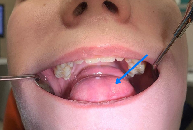
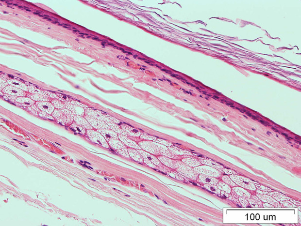
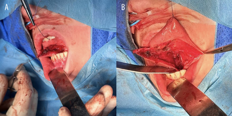
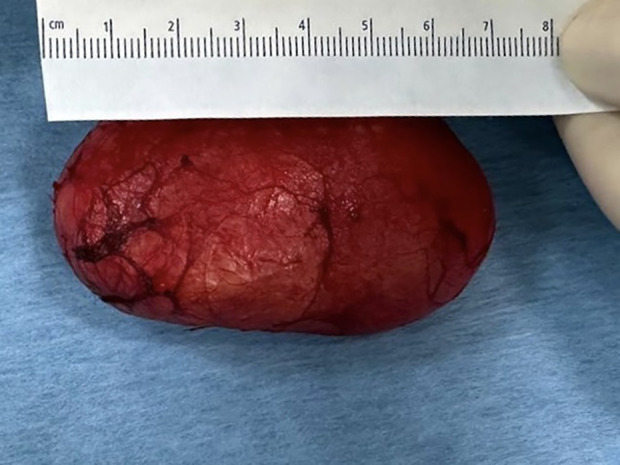
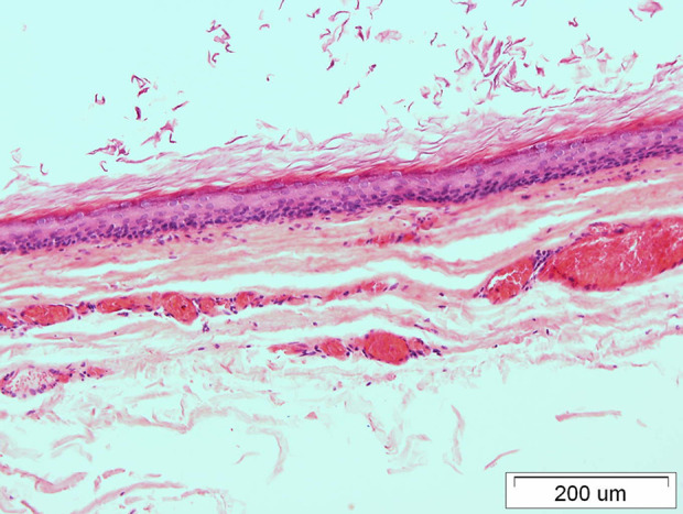
BACKGROUND Ranulas are typical causes of sublingual cysts in children. However, our case was histopathologically confirmed to be a dermoid cyst. Epidermoid and dermoid cysts of the floor of the mouth account for <0.01% of all oral cavity lesions and 0.29% of head and neck tumors in children. Salivary congestive cysts (ranulas) involve the sublingual salivary glands or the common duct of the sublingual and submandibular salivary glands. This report describes a 13-year-old boy presenting with a sublingual cyst, diagnosed by histopathology as a dermoid cyst. Treatment is based on surgical removal of the cyst, sometimes along with the altered salivary gland. CASE REPORT A 13-year-old boy was admitted to the Department of Otolaryngology with the Subdivision of Maxillofacial Surgery for the diagnosis of a tumor localized under the tongue. A significant growth of the tumor during a 3-month period was noticed, with appearance of a mass effect, speech disorders, and difficulties in eating. Significant elevation of the floor of the mouth and tongue was shown. The presence a ranula was indicated. Surgical excision was performed using intra-oral excision. Histopathological examination revealed a diagnosis of dermatoid cyst. CONCLUSIONS This case highlights the importance of detailed histopathological diagnosis of lesions and the usefulness of imaging methods like magnetic resonance imaging (MRI), ultrasound (US) or computed tomography (CT). Our patient had a dermoid cyst, which appears rarely among children in the floor of the mouth. This shows the significance of their proper differentiation, as some may be misdiagnosed as ranula.
背景 舌下囊肿是儿童舌下囊肿的典型病因。然而,我们的病例经组织病理学证实为皮样囊肿。口腔底部的表皮样囊肿和皮样囊肿占所有口腔病变的比例<0.01%,占儿童头颈部肿瘤的0.29%。唾液潴留性囊肿(舌下囊肿)累及舌下腺或舌下腺与下颌下腺的共同导管。本报告描述了一名13岁男孩,表现为舌下囊肿,经组织病理学诊断为皮样囊肿。治疗基于手术切除囊肿,有时连同病变的唾液腺一并切除。病例报告 一名13岁男孩因诊断舌下肿物入住耳鼻咽喉科颌面外科亚专业。在3个月期间发现肿物显著生长,出现占位效应、言语障碍和进食困难。可见口底和舌明显抬高。提示存在舌下囊肿。采用口内切除术进行手术切除。组织病理学检查确诊为皮样囊肿。结论 本病例强调了病变详细组织病理学诊断的重要性以及磁共振成像(MRI)、超声(US)或计算机断层扫描(CT)等影像学方法的实用性。我们的患者患有皮样囊肿,在儿童口腔底部较为罕见。这显示了正确鉴别它们的重要性,因为有些可能会被误诊为舌下囊肿。