• 文献检索
  • 文档翻译
  • 深度研究
  • 学术资讯
  • Suppr Zotero 插件Zotero 插件
  • 邀请有礼
  • 套餐&价格
  • 历史记录
应用&插件
Suppr Zotero 插件Zotero 插件浏览器插件Mac 客户端Windows 客户端微信小程序
定价
高级版会员购买积分包购买API积分包
服务
文献检索文档翻译深度研究API 文档MCP 服务
关于我们
关于 Suppr公司介绍联系我们用户协议隐私条款
关注我们

Suppr 超能文献

核心技术专利:CN118964589B侵权必究
粤ICP备2023148730 号-1Suppr @ 2026

文献检索

告别复杂PubMed语法,用中文像聊天一样搜索,搜遍4000万医学文献。AI智能推荐,让科研检索更轻松。

立即免费搜索

文件翻译

保留排版,准确专业,支持PDF/Word/PPT等文件格式,支持 12+语言互译。

免费翻译文档

深度研究

AI帮你快速写综述,25分钟生成高质量综述,智能提取关键信息,辅助科研写作。

立即免费体验

外侧开口楔形股骨远端截骨术中保护克氏针直径的定量分析:一项有限元研究

Quantitative analysis of protective Kirschner wire diameters in lateral opening wedge distal femoral osteotomy: A finite element study.

作者信息

Baris Alican, Özmen Emre, Circi Esra, Yuksel Serdar, Beytemür Ozan

机构信息

İstanbul Fizik Tedavi ve Rehabilitasyon Eğitim ve Araştırma Hastanesi, Ortopedi ve Travmatoloji Kliniği, 34180 Bahçelievler, İstanbul, Türkiye.

出版信息

Jt Dis Relat Surg. 2025 Jan 2;36(1):97-106. doi: 10.52312/jdrs.2025.1806. Epub 2024 Nov 5.

DOI:10.52312/jdrs.2025.1806
PMID:39719906
原文链接:https://pmc.ncbi.nlm.nih.gov/articles/PMC11734846/
Abstract

OBJECTIVES

This study aims to investigate quantitatively the protective effect of a 1.6-mm or a 2.5-mm Kirschner wire (K-wire) on the medial hinge at different gap distances through finite element analysis (FEA) and to establish whether using a 2.5-mm K-wire can offer benefits compared to a 1.6-mm in preventing medial hinge fractures.

MATERIALS AND METHODS

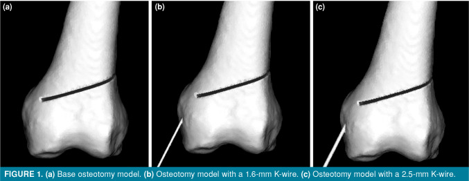
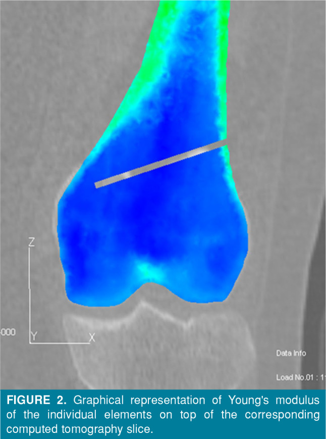
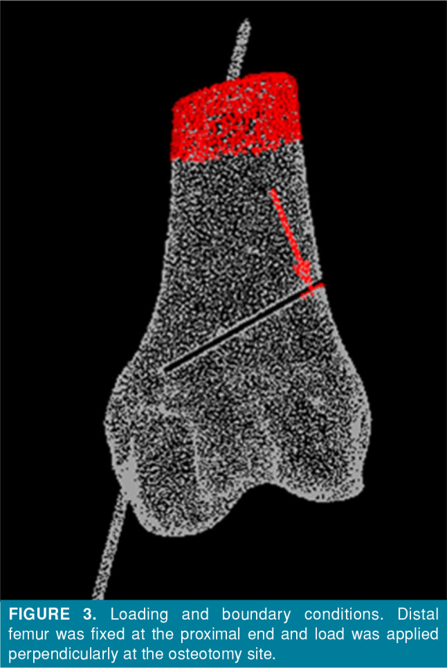
Between June 2024 and July 2024, three different models simulating a lateral opening wedge (LOW) osteotomy of the distal femur were created from a femoral computed tomography (CT) scan of a 36-year-old male patient: no K-wire (Model I), 1.6-mm K-wire (Model II), and 2.5-mm K-wire (Model III). Finite element analysis was performed to simulate 7- to 13-mm gaps at the osteotomy site. Loads, principal stress, strain, and equivalent stress were analyzed around the medial hinge.

RESULTS

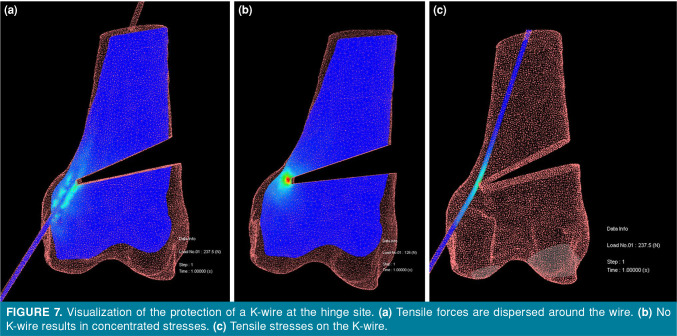
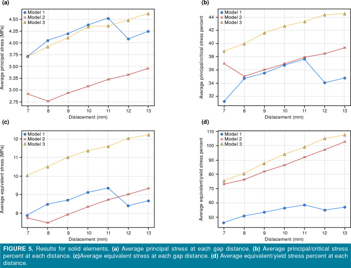
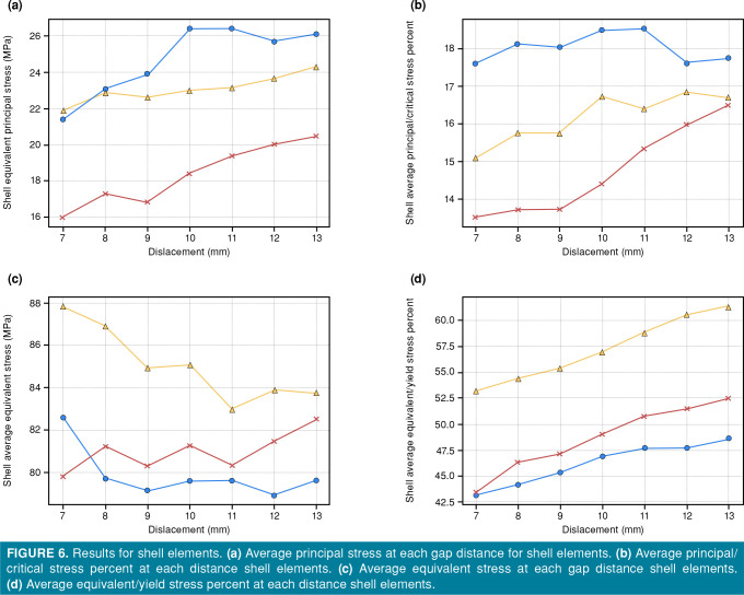
Model I required 123.0±5.2 N, Model II required 181.7±12.2 N, and Model III required 228.7±13.6 N (p<0.001). Cracked shell elements were the lowest in Model II and the highest in Model I. While the average equivalent/yield stress ratio was not significantly lower in Model II compared to Model III (87.0±10.9% vs. 92.7±12.1%), the maximum equivalent/yield stress ratio values in Model II were significantly lower than both Model I and Model III (1206.2±138.3% vs. 1836.2±165.4% and 1689.1±404.0%, respectively), suggesting a superior dispersion of forces.

CONCLUSION

Using a 1.6-mm K-wire during LOW osteotomy of the distal femur provides a balance between structural reinforcement and stress distribution, significantly improving stability and reducing the risk of medial hinge fractures compared to a 2.5-mm K-wire or no K-wire. The 1.6-mm K-wire optimizes stress dispersion, making it the preferred choice for surgical planning in lateral opening wedge distal femoral osteotomy.

摘要

目的

本研究旨在通过有限元分析(FEA)定量研究1.6毫米或2.5毫米克氏针(K针)在不同间隙距离下对内侧铰链的保护作用,并确定与1.6毫米K针相比,使用2.5毫米K针在预防内侧铰链骨折方面是否具有优势。

材料与方法

在2024年6月至2024年7月期间,从一名36岁男性患者的股骨计算机断层扫描(CT)扫描中创建了三种不同的模型,模拟股骨远端外侧开口楔形(LOW)截骨术:无K针(模型I)、1.6毫米K针(模型II)和2.5毫米K针(模型III)。进行有限元分析以模拟截骨部位7至13毫米的间隙。分析内侧铰链周围的载荷、主应力、应变和等效应力。

结果

模型I需要123.0±5.2 N,模型II需要181.7±12.2 N,模型III需要228.7±13.6 N(p<0.001)。破裂的壳单元在模型II中最低,在模型I中最高。虽然模型II的平均等效应力/屈服应力比与模型III相比没有显著降低(87.0±10.9%对92.7±12.1%),但模型II中的最大等效应力/屈服应力比值显著低于模型I和模型III(分别为1206.2±138.3%对1836.2±165.4%和1689.1±404.0%),表明力的分散性更好。

结论

在股骨远端LOW截骨术中使用1.6毫米K针可在结构加固和应力分布之间取得平衡,与2.5毫米K针或不使用K针相比,显著提高稳定性并降低内侧铰链骨折的风险。1.6毫米K针优化了应力分散,使其成为股骨远端外侧开口楔形截骨术手术规划的首选。

https://cdn.ncbi.nlm.nih.gov/pmc/blobs/d60c/11734846/0eb5bbf92c35/JDRS-2025-36-1-097-106-F7.jpg
https://cdn.ncbi.nlm.nih.gov/pmc/blobs/d60c/11734846/581e91807187/JDRS-2025-36-1-097-106-F1.jpg
https://cdn.ncbi.nlm.nih.gov/pmc/blobs/d60c/11734846/80dfa1960bd3/JDRS-2025-36-1-097-106-F2.jpg
https://cdn.ncbi.nlm.nih.gov/pmc/blobs/d60c/11734846/cb9fc774eed1/JDRS-2025-36-1-097-106-F3.jpg
https://cdn.ncbi.nlm.nih.gov/pmc/blobs/d60c/11734846/8ce4ecaea363/JDRS-2025-36-1-097-106-F4.jpg
https://cdn.ncbi.nlm.nih.gov/pmc/blobs/d60c/11734846/eb7a7bdb823f/JDRS-2025-36-1-097-106-F5.jpg
https://cdn.ncbi.nlm.nih.gov/pmc/blobs/d60c/11734846/2861753ca1b4/JDRS-2025-36-1-097-106-F6.jpg
https://cdn.ncbi.nlm.nih.gov/pmc/blobs/d60c/11734846/0eb5bbf92c35/JDRS-2025-36-1-097-106-F7.jpg

相似文献

1
Quantitative analysis of protective Kirschner wire diameters in lateral opening wedge distal femoral osteotomy: A finite element study.外侧开口楔形股骨远端截骨术中保护克氏针直径的定量分析:一项有限元研究
Jt Dis Relat Surg. 2025 Jan 2;36(1):97-106. doi: 10.52312/jdrs.2025.1806. Epub 2024 Nov 5.
2
The Ideal Location of the Lateral Hinge in Medial Closing Wedge Osteotomy of the Distal Femur: Analysis of Soft Tissue Coverage and Bone Density.外侧铰链在股骨远端内侧闭合楔形截骨术中的理想位置:软组织覆盖和骨密度分析。
Am J Sports Med. 2019 Oct;47(12):2945-2951. doi: 10.1177/0363546519869325. Epub 2019 Aug 29.
3
Adding a protective K-wire during opening high tibial osteotomy increases lateral hinge resistance to fracture.在胫骨高位截骨术中增加保护性 K 线可增加外侧铰链抵抗骨折的能力。
Knee Surg Sports Traumatol Arthrosc. 2020 Mar;28(3):751-758. doi: 10.1007/s00167-019-05404-7. Epub 2019 Feb 19.
4
Finite Element Analysis of Protective Measures against Lateral Hinge Fractures in High-Tibial Osteotomy.胫骨高位截骨术中防止外侧铰链骨折的保护措施的有限元分析
Adv Orthop. 2024 Aug 22;2024:5510319. doi: 10.1155/2024/5510319. eCollection 2024.
5
A protective hinge wire, intersecting the osteotomy plane, can reduce the occurrence of perioperative hinge fractures in medial opening wedge osteotomy.保护性铰链钢丝,与截骨面相交,可以减少内侧撑开截骨术中铰链骨折的发生。
Knee Surg Sports Traumatol Arthrosc. 2020 Oct;28(10):3173-3182. doi: 10.1007/s00167-019-05806-7. Epub 2019 Nov 26.
6
Impact of support instruments in medial closed-wedge distal femoral osteotomy: a finite element analysis.支撑器械对内侧闭合楔形股骨远端截骨术的影响:有限元分析。
Arch Orthop Trauma Surg. 2024 Mar;144(3):1039-1045. doi: 10.1007/s00402-023-05152-x. Epub 2023 Dec 19.
7
Additional Plate Fixation of Hinge Fractures After Varisation Distal Femoral Osteotomies Provides Favorable Torsional Stability: A Biomechanical Study.铰链骨折术后附加钢板固定可提供良好的扭转稳定性:一项生物力学研究。
Am J Sports Med. 2023 Dec;51(14):3732-3741. doi: 10.1177/03635465231206947. Epub 2023 Nov 7.
8
Experimental study of risk of medial hinge fracture during distal femoral varus osteotomy.股骨远端内翻截骨术中内侧铰链骨折风险的实验研究。
Orthop Traumatol Surg Res. 2023 Jun;109(4):103527. doi: 10.1016/j.otsr.2022.103527. Epub 2022 Dec 20.
9
The effect of the angle between fracture line and Kirschner wires on stability in supracondylar humerus fractures treated with Kirschner wire fixation: A finite element analysis.骨折线与克氏针之间角度对克氏针固定治疗肱骨髁上骨折稳定性的影响:有限元分析。
Jt Dis Relat Surg. 2021;32(1):75-84. doi: 10.5606/ehc.2021.77279. Epub 2021 Jan 6.
10
Biomechanical effect of a lateral hinge fracture for a medial opening wedge high tibial osteotomy: finite element study.内侧开口楔形胫骨高位截骨术横向铰链骨折的生物力学效应:有限元研究。
J Orthop Surg Res. 2020 Feb 21;15(1):63. doi: 10.1186/s13018-020-01597-7.

本文引用的文献

1
Effects of preoperative valgus deformity in patients undergoing neutrally aligned total knee arthroplasty: A retrospective cohort study with a minimum five-year follow-up.接受中立对线全膝关节置换术的患者术前外翻畸形的影响:一项最少随访 5 年的回顾性队列研究。
Jt Dis Relat Surg. 2024 Aug 14;35(3):529-537. doi: 10.52312/jdrs.2024.1800.
2
Simultaneous medial closing wedge distal femoral varus osteotomy and double-bundle anterior cruciate ligament reconstruction in the symptomatic femoral valgus deformity: A case report.症状性股骨外翻畸形中同时行内侧闭合楔形股骨远端内翻截骨术和双束前交叉韧带重建术:病例报告。
Jt Dis Relat Surg. 2024 Feb 13;35(2):422-432. doi: 10.52312/jdrs.2023.1176.
3
Impact of support instruments in medial closed-wedge distal femoral osteotomy: a finite element analysis.
支撑器械对内侧闭合楔形股骨远端截骨术的影响:有限元分析。
Arch Orthop Trauma Surg. 2024 Mar;144(3):1039-1045. doi: 10.1007/s00402-023-05152-x. Epub 2023 Dec 19.
4
Writing for Joint Diseases and Related Surgery (JDRS): There is something new and interesting in this article!为《关节疾病与相关外科手术》(JDRS)撰写文章:本文有一些新颖有趣的内容!
Jt Dis Relat Surg. 2023 Sep 21;34(3):533. doi: 10.52312/jdrs.2023.57916.
5
Finite element analysis of medial closing and lateral opening wedge osteotomies of the distal femur in relation to hinge fractures.股骨远端内侧闭合和外侧开放楔形截骨术与铰链骨折相关的有限元分析
J Exp Orthop. 2023 Mar 27;10(1):33. doi: 10.1186/s40634-023-00597-w.
6
Large Versus Small Opening Wedge High Tibial Osteotomies Performed With a Protective Wire Over the Lateral Hinge: Incidence of Lateral Hinge Fracture and Early Clinical Outcomes.在外侧铰链处使用保护钢丝进行的大开口与小开口高位胫骨截骨术:外侧铰链骨折的发生率及早期临床结果
Am J Sports Med. 2023 Mar;51(3):672-677. doi: 10.1177/03635465221148496.
7
Experimental study of risk of medial hinge fracture during distal femoral varus osteotomy.股骨远端内翻截骨术中内侧铰链骨折风险的实验研究。
Orthop Traumatol Surg Res. 2023 Jun;109(4):103527. doi: 10.1016/j.otsr.2022.103527. Epub 2022 Dec 20.
8
Distalization of hinge site with use of hinge wire reduces hinge fracture rates in closing wedge distal femoral osteotomy.使用铰链线远移铰链部位可降低闭合楔形股骨远端截骨术中铰链断裂的发生率。
Knee Surg Sports Traumatol Arthrosc. 2023 Aug;31(8):3141-3150. doi: 10.1007/s00167-022-07286-8. Epub 2022 Dec 23.
9
Experimental investigation of the risk of lateral cortex fracture during valgus tibial osteotomy.外侧皮质骨折风险在胫骨外翻截骨术中的实验研究。
Orthop Traumatol Surg Res. 2022 Dec;108(8):103428. doi: 10.1016/j.otsr.2022.103428. Epub 2022 Oct 3.
10
Hinge fractures reaching the tibial plateau can be caused by forcible opening of insufficient posterior osteotomy during open-wedge high tibial osteotomy.在开放性楔形高位胫骨截骨术中,到达胫骨平台的铰链骨折可能是由于后截骨不足时强行打开所致。
Knee Surg Sports Traumatol Arthrosc. 2023 Apr;31(4):1533-1545. doi: 10.1007/s00167-021-06816-0. Epub 2022 Jan 4.