Boulestreau Jérémy, Molina Laurence, Ouedraogo Alimata, Laramy Louën, Grich Ines, Van Thi Nhu Ngoc, Molina Franck, Kahli Malik
Sys2Diag, UMR9005 CNRS/ALCEN, Cap Gamma, Parc Euromédecine, 1682 Rue de la Valsière, CS 40182, 34184, Montpellier Cedex 4, France.
Department of Anatomy, Biochemistry, and Physiology John A. Burns School of Medicine, University of Hawaii at Manoa, 651 Ilalo St. BSB 211, Honolulu, HI, 96813, USA.
Sci Rep. 2024 Dec 28;14(1):31233. doi: 10.1038/s41598-024-82488-3.
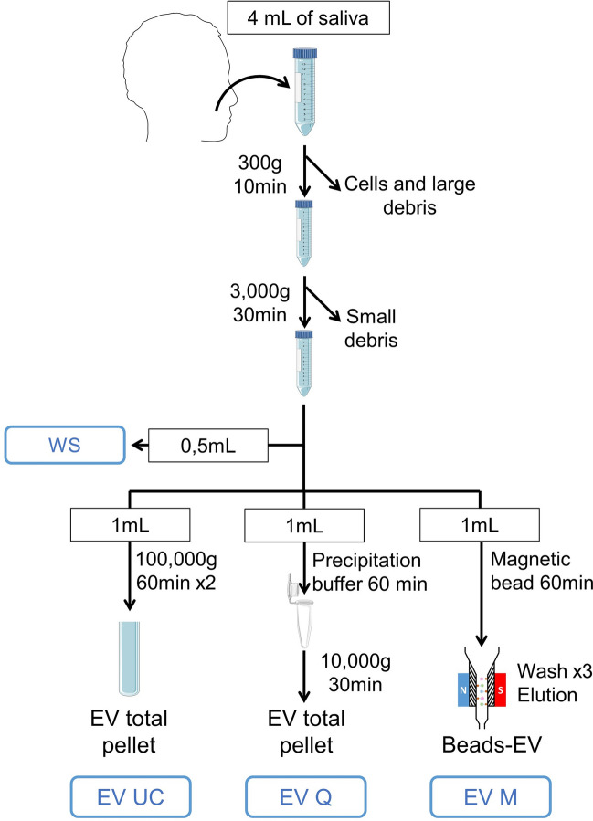
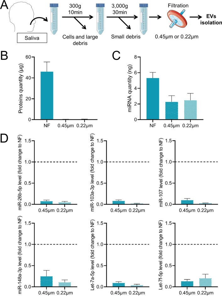
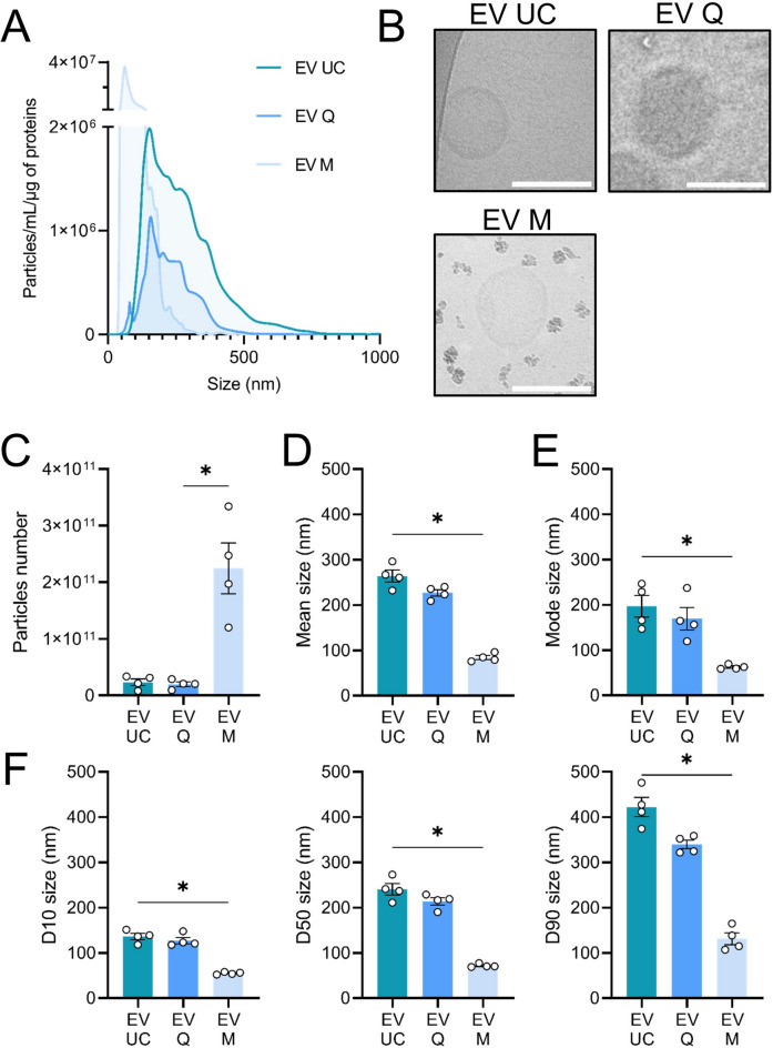
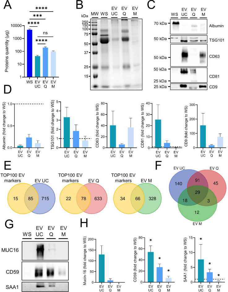
Extracellular vesicles (EVs), crucial mediators in cell-to-cell communication, are implicated in both homeostatic and pathological processes. Their detectability in easily accessible peripheral fluids like saliva positions them as promising candidates for non-invasive biomarker discovery. However, the lack of standardized methods for salivary EVs isolation greatly limits our ability to study them. Therefore, we rigorously compared salivary EVs isolated using two scalable techniques-co-precipitation and immuno-affinity-against the long-established but labor-intensive ultracentrifugation method. Employing Cryo-Electron Microscopy (Cryo-EM), Nanoparticle Tracking Analysis, Western blots (WB), and proteomics, we identified significant method-dependent variances in the size, concentration, and protein content of EVs. Importantly, our study uniquely demonstrates the ability of EV isolation to detect specific biomarkers that remain undetected in whole saliva by WB. RT-qPCR analysis targeting six miRNAs confirmed a consistent enrichment of these miRNAs in EV-derived cargo across all three isolation methods. We also found that pre-filtering saliva samples with 0.22 or 0.45 µm pores adversely affects subsequent analyses. Our findings highlight the untapped potential of salivary EVs in diagnostics and advocate for the co-precipitation method as an efficient, cost-effective, and clinically relevant approach for small-volume saliva samples. This work not only sheds light on a neglected source of EVs but also paves the way for their application in routine clinical diagnostics.
细胞外囊泡(EVs)是细胞间通讯的关键介质,参与稳态和病理过程。它们在唾液等易于获取的外周液中的可检测性使其成为非侵入性生物标志物发现的有希望的候选者。然而,缺乏标准化的唾液EVs分离方法极大地限制了我们对其进行研究的能力。因此,我们严格比较了使用两种可扩展技术——共沉淀和免疫亲和——分离的唾液EVs与已确立但劳动强度大的超速离心法。通过冷冻电子显微镜(Cryo-EM)、纳米颗粒跟踪分析、蛋白质印迹(WB)和蛋白质组学,我们确定了EVs在大小、浓度和蛋白质含量方面存在显著的方法依赖性差异。重要的是,我们的研究独特地证明了EVs分离能够检测到WB在全唾液中未检测到的特定生物标志物。针对六种miRNA的RT-qPCR分析证实,在所有三种分离方法中,这些miRNA在EV衍生的货物中均持续富集。我们还发现,用0.22或0.45 µm孔径的滤膜对唾液样本进行预过滤会对后续分析产生不利影响。我们的研究结果突出了唾液EVs在诊断方面尚未开发的潜力,并提倡将共沉淀法作为一种高效、经济且与临床相关的方法用于小体积唾液样本。这项工作不仅揭示了一个被忽视的EVs来源,也为其在常规临床诊断中的应用铺平了道路。