Qiao Qincheng, Cao Juan, Xue Wen, Qian Jin, Wang Chuan, Pan Qi, Lu Bin, Xiong Qian, Chen Li, Hou Xinguo
Department of Endocrinology and Metabolism, Qilu Hospital, Shandong University, Jinan, China.
The First Clinical Medical College, Cheeloo College of Medicine, Shandong University, Jinan, China.
Digit Health. 2024 Dec 25;10:20552076241307573. doi: 10.1177/20552076241307573. eCollection 2024 Jan-Dec.
Diabetic peripheral neuropathy (DPN) is a common complication of diabetes, and its early identification is crucial for improving patient outcomes. Corneal confocal microscopy (CCM) can non-invasively detect changes in corneal nerve fibers (CNFs), making it a potential tool for the early diagnosis of DPN. However, the existing CNF analysis methods have certain limitations, highlighting the need to develop a reliable automated analysis tool.
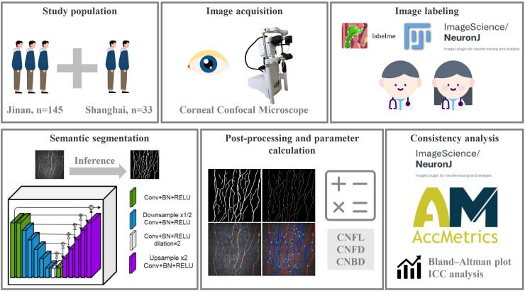
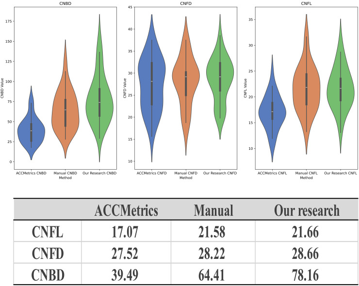
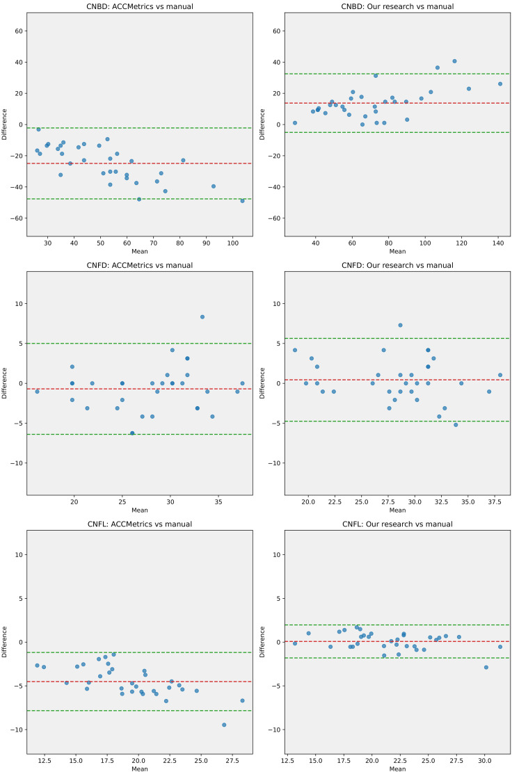
This study is based on data from two independent clinical centers. Various popular deep learning (DL) models have been trained and evaluated for their performance in CCM image segmentation using DL-based image segmentation techniques. Subsequently, an image processing algorithm was designed to automatically extract and quantify various morphological parameters of CNFs. To validate the effectiveness of this tool, it was compared with manually annotated datasets and ACCMetrics, and the consistency of the results was assessed using Bland--Altman analysis and intraclass correlation coefficient (ICC).
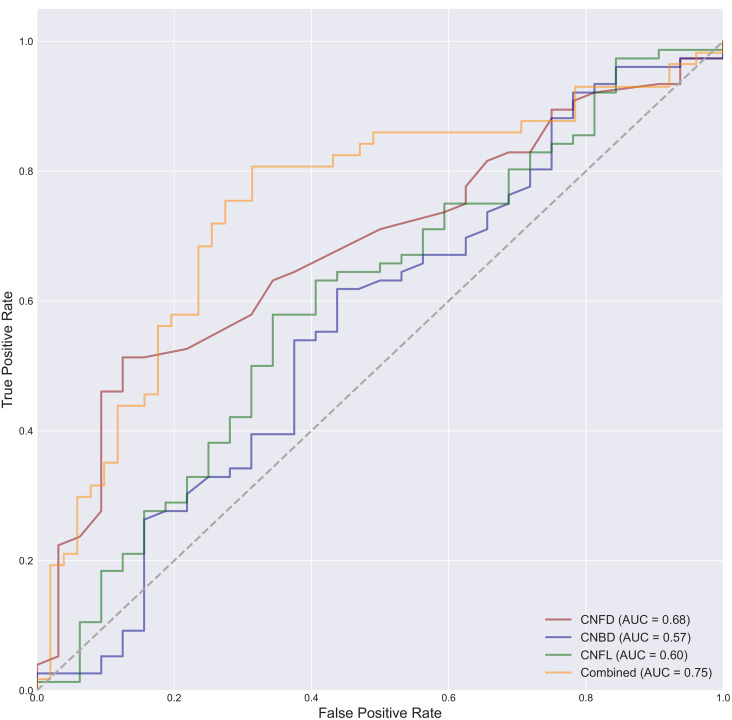
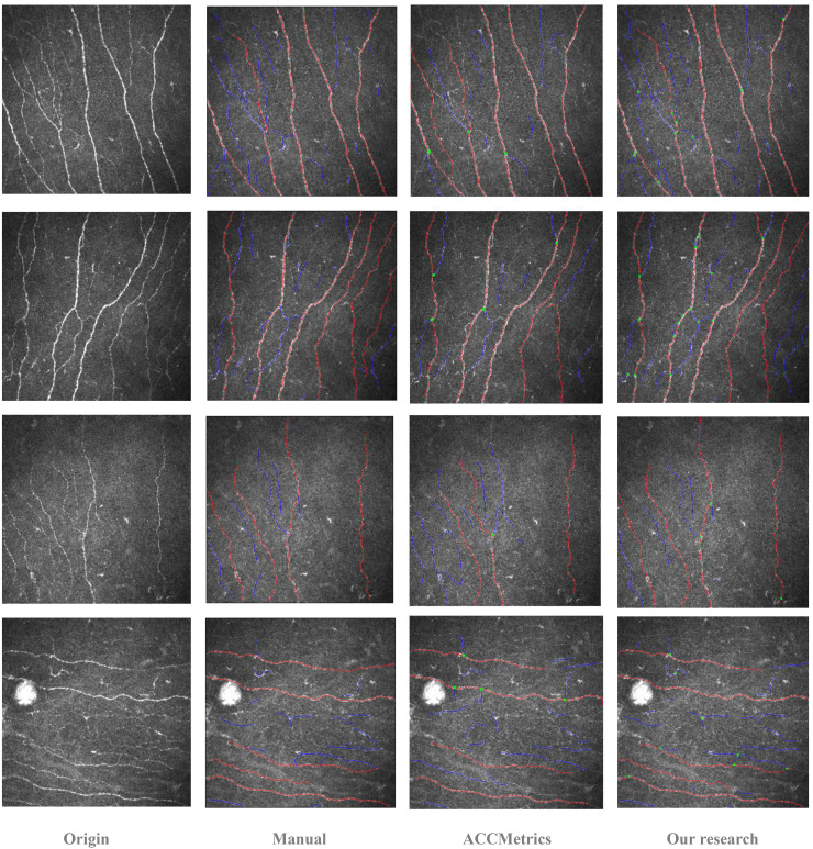
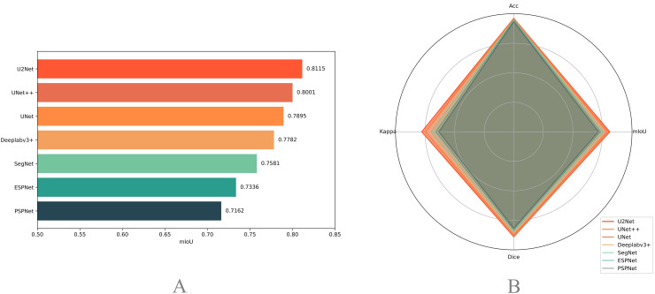
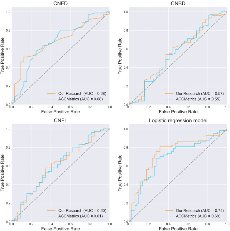
The U2Net model performed the best in the CCM image segmentation task, achieving a mean Intersection over Union (mIoU) of 0.8115. The automated analysis tool based on U2Net demonstrated a significantly higher consistency with the manually annotated results in the quantitative analysis of various CNF morphological parameters than the previously popular automated tool ACCMetrics. The area under the curve for classifying DPN using the CNF morphology parameters calculated by this tool reached 0.75.
The DL-based automated tool developed in this study can effectively segment and quantify the CNF parameters in CCM images. This tool has the potential to be used for the early diagnosis of DPN, and further research will help validate its practical application value in clinical settings.
糖尿病周围神经病变(DPN)是糖尿病常见的并发症,早期识别对于改善患者预后至关重要。角膜共焦显微镜检查(CCM)可无创检测角膜神经纤维(CNF)的变化,使其成为DPN早期诊断的潜在工具。然而,现有的CNF分析方法存在一定局限性,凸显了开发可靠的自动化分析工具的必要性。
本研究基于来自两个独立临床中心的数据。使用基于深度学习的图像分割技术,对各种流行的深度学习(DL)模型进行了训练,并评估了它们在CCM图像分割中的性能。随后,设计了一种图像处理算法,以自动提取和量化CNF的各种形态学参数。为验证该工具的有效性,将其与手动标注的数据集和ACCMetrics进行比较,并使用布兰德-奥特曼分析和组内相关系数(ICC)评估结果的一致性。
U2Net模型在CCM图像分割任务中表现最佳,平均交并比(mIoU)达到0.8115。基于U2Net的自动化分析工具在各种CNF形态学参数的定量分析中,与手动标注结果的一致性显著高于先前流行的自动化工具ACCMetrics。使用该工具计算的CNF形态学参数对DPN进行分类的曲线下面积达到0.75。
本研究开发的基于深度学习的自动化工具能够有效分割和量化CCM图像中的CNF参数。该工具具有用于DPN早期诊断的潜力,进一步的研究将有助于验证其在临床环境中的实际应用价值。