• 文献检索
  • 文档翻译
  • 深度研究
  • 学术资讯
  • Suppr Zotero 插件Zotero 插件
  • 邀请有礼
  • 套餐&价格
  • 历史记录
应用&插件
Suppr Zotero 插件Zotero 插件浏览器插件Mac 客户端Windows 客户端微信小程序
定价
高级版会员购买积分包购买API积分包
服务
文献检索文档翻译深度研究API 文档MCP 服务
关于我们
关于 Suppr公司介绍联系我们用户协议隐私条款
关注我们

Suppr 超能文献

核心技术专利:CN118964589B侵权必究
粤ICP备2023148730 号-1Suppr @ 2026

文献检索

告别复杂PubMed语法,用中文像聊天一样搜索,搜遍4000万医学文献。AI智能推荐,让科研检索更轻松。

立即免费搜索

文件翻译

保留排版,准确专业,支持PDF/Word/PPT等文件格式,支持 12+语言互译。

免费翻译文档

深度研究

AI帮你快速写综述,25分钟生成高质量综述,智能提取关键信息,辅助科研写作。

立即免费体验

使用卷积神经网络的自动化程序用于角膜共焦显微镜图像的客观且可重复选择。

Automated program using convolutional neural networks for objective and reproducible selection of corneal confocal microscopy images.

作者信息

Qiao Qincheng, Xue Wen, Li Jinzhe, Zheng Wenwen, Yuan Yongkai, Li Chen, Liu Fuqiang, Hou Xinguo

机构信息

Department of Endocrinology and Metabolism, Qilu Hospital, Shandong University, Jinan, China.

The First Clinical Medical College, Cheeloo College of Medicine, Shandong University, Jinan, China.

出版信息

Digit Health. 2025 Mar 17;11:20552076251326223. doi: 10.1177/20552076251326223. eCollection 2025 Jan-Dec.

DOI:10.1177/20552076251326223
PMID:40103638
原文链接:https://pmc.ncbi.nlm.nih.gov/articles/PMC11915551/
Abstract

OBJECTIVE

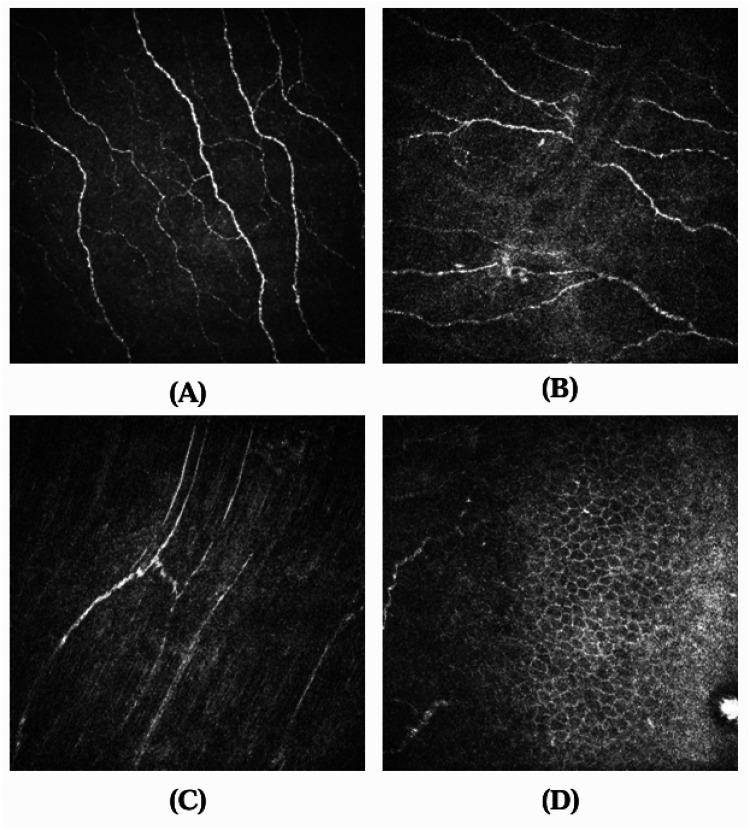
Diabetic peripheral neuropathy (DPN) is a common complication of diabetes, posing a significant risk for foot ulcers and amputation. Corneal confocal microscopy (CCM) is a rapid, noninvasive method to assess DPN by analysing corneal nerve fibre morphology. However, selecting high-quality representative images remains a critical challenge.

METHODS

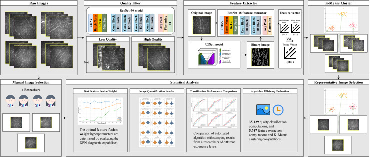
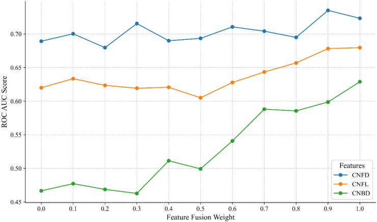
In this study, we propose a fully automated CCM image-selection algorithm based on deep learning feature extraction using ResNet-18 and unsupervised clustering. The algorithm consistently identifies representative images by balancing non-redundancy and representativeness, ensuring objectivity and reproducibility.

RESULTS

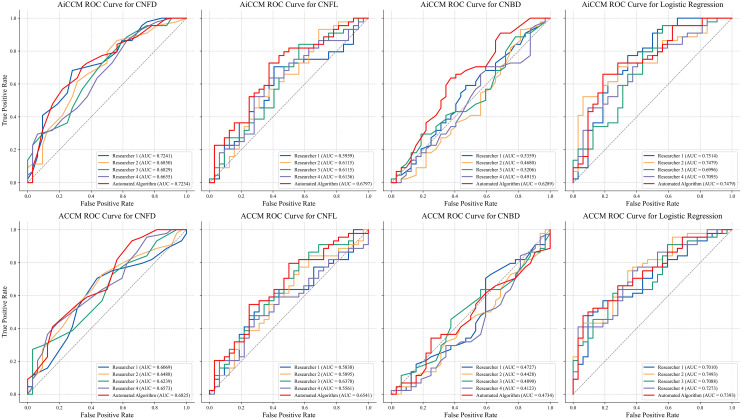
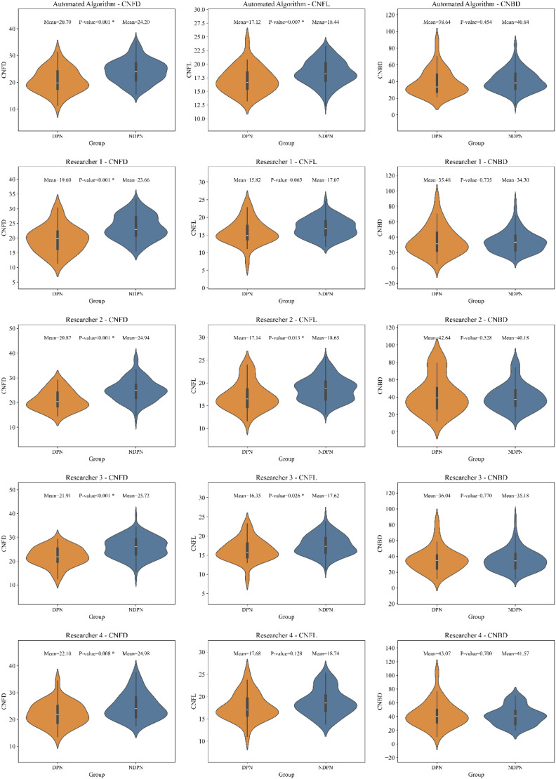
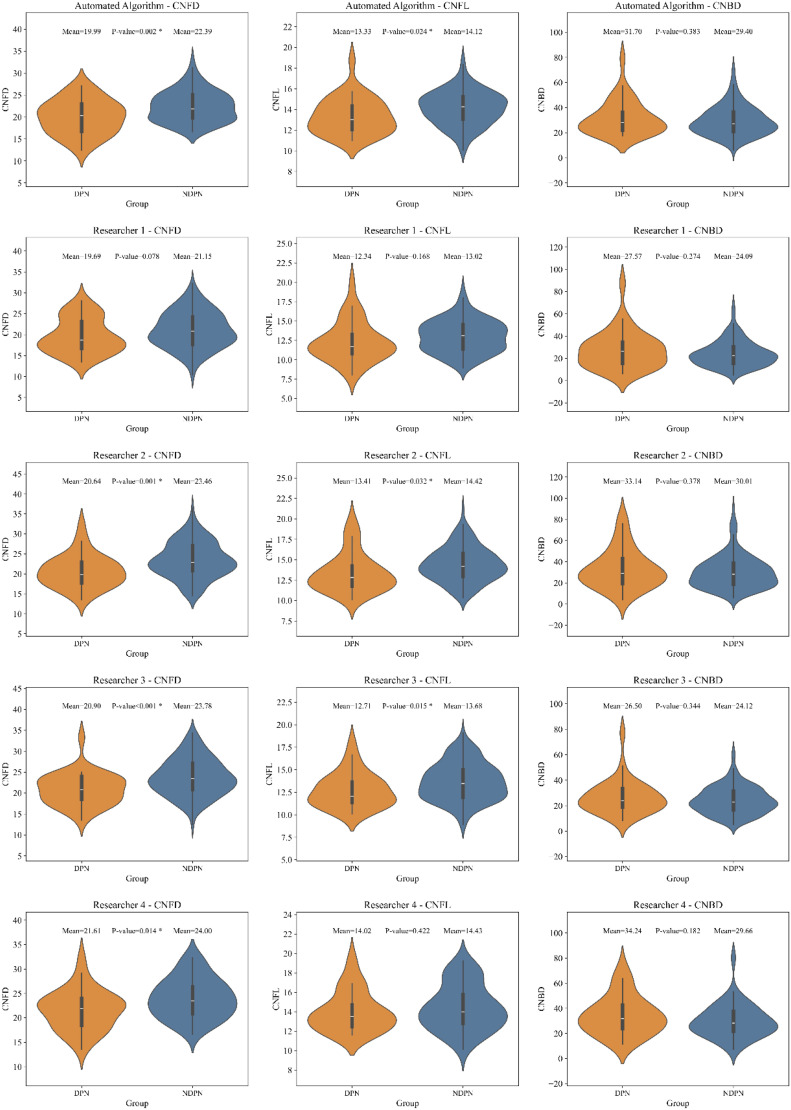
When validated against manual selection by researchers with varying expertise levels, the algorithm demonstrated superior performance in distinguishing DPN and reduced inter-observer variability. It completed the analysis of hundreds of images within 1 s, significantly enhancing diagnostic efficiency. Compared with traditional manual selection, the proposed method achieved higher diagnostic accuracy for key morphological parameters, including corneal nerve fibre density, length, and branch density.

CONCLUSION

The algorithm is open source and compatible with standard CCM workflows, offering researchers and clinicians a robust and efficient tool for DPN diagnosis. Further, multicentre studies are needed to validate these findings in diverse populations.

摘要

目的

糖尿病周围神经病变(DPN)是糖尿病常见的并发症,会引发足部溃疡和截肢的重大风险。角膜共焦显微镜检查(CCM)是一种通过分析角膜神经纤维形态来评估DPN的快速、非侵入性方法。然而,选择高质量的代表性图像仍然是一个关键挑战。

方法

在本研究中,我们提出了一种基于深度学习特征提取(使用ResNet - 18)和无监督聚类的全自动CCM图像选择算法。该算法通过平衡非冗余性和代表性来持续识别代表性图像,确保客观性和可重复性。

结果

在与不同专业水平的研究人员进行手动选择验证时,该算法在区分DPN方面表现出卓越性能,并减少了观察者间的变异性。它在1秒内完成了数百张图像的分析,显著提高了诊断效率。与传统手动选择相比,该方法在关键形态学参数(包括角膜神经纤维密度、长度和分支密度)方面实现了更高的诊断准确性。

结论

该算法是开源的,并且与标准CCM工作流程兼容,为研究人员和临床医生提供了一个用于DPN诊断的强大且高效的工具。此外,需要进行多中心研究以在不同人群中验证这些发现。

https://cdn.ncbi.nlm.nih.gov/pmc/blobs/afb1/11915551/f8bf74a24787/10.1177_20552076251326223-fig6.jpg
https://cdn.ncbi.nlm.nih.gov/pmc/blobs/afb1/11915551/f0d6ed416263/10.1177_20552076251326223-fig1.jpg
https://cdn.ncbi.nlm.nih.gov/pmc/blobs/afb1/11915551/9409092ba721/10.1177_20552076251326223-fig2.jpg
https://cdn.ncbi.nlm.nih.gov/pmc/blobs/afb1/11915551/9dde7105bbcc/10.1177_20552076251326223-fig3.jpg
https://cdn.ncbi.nlm.nih.gov/pmc/blobs/afb1/11915551/f2797ad54cbd/10.1177_20552076251326223-fig4.jpg
https://cdn.ncbi.nlm.nih.gov/pmc/blobs/afb1/11915551/52f56308b5d9/10.1177_20552076251326223-fig5.jpg
https://cdn.ncbi.nlm.nih.gov/pmc/blobs/afb1/11915551/f8bf74a24787/10.1177_20552076251326223-fig6.jpg

相似文献

1
Automated program using convolutional neural networks for objective and reproducible selection of corneal confocal microscopy images.使用卷积神经网络的自动化程序用于角膜共焦显微镜图像的客观且可重复选择。
Digit Health. 2025 Mar 17;11:20552076251326223. doi: 10.1177/20552076251326223. eCollection 2025 Jan-Dec.
2
An artificial intelligence-based deep learning algorithm for the diagnosis of diabetic neuropathy using corneal confocal microscopy: a development and validation study.基于人工智能的深度学习算法利用角膜共聚焦显微镜诊断糖尿病周围神经病变:一项开发和验证研究。
Diabetologia. 2020 Feb;63(2):419-430. doi: 10.1007/s00125-019-05023-4. Epub 2019 Nov 12.
3
Artificial Intelligence and Corneal Confocal Microscopy: The Start of a Beautiful Relationship.人工智能与角膜共焦显微镜:美好关系的开端。
J Clin Med. 2022 Oct 20;11(20):6199. doi: 10.3390/jcm11206199.
4
Corneal confocal microscopy for the diagnosis of diabetic peripheral neuropathy: A systematic review and meta-analysis.角膜共焦显微镜在糖尿病周围神经病变诊断中的应用:系统评价和荟萃分析。
J Diabetes Investig. 2022 Jan;13(1):134-147. doi: 10.1111/jdi.13643. Epub 2021 Aug 27.
5
Development of a transformer-based deep learning algorithm for diabetic peripheral neuropathy classification using corneal confocal microscopy images.基于变压器的深度学习算法用于利用角膜共聚焦显微镜图像进行糖尿病周围神经病变分类的开发。
Front Cell Dev Biol. 2024 Oct 14;12:1484329. doi: 10.3389/fcell.2024.1484329. eCollection 2024.
6
Utility of corneal confocal microscopy for assessing mild diabetic neuropathy: baseline findings of the LANDMark study.角膜共焦显微镜在评估轻度糖尿病神经病变中的应用:LANDMark研究的基线结果
Clin Exp Optom. 2012 May;95(3):348-54. doi: 10.1111/j.1444-0938.2012.00740.x. Epub 2012 Apr 29.
7
Improved sampling and analysis of images in corneal confocal microscopy.角膜共焦显微镜术中图像采样与分析的改进
J Microsc. 2017 Oct;268(1):3-12. doi: 10.1111/jmi.12581. Epub 2017 May 26.
8
Mosaic vs. Single Image Analysis with Confocal Microscopy of the Corneal Nerve Plexus for Diagnosis of Early Diabetic Peripheral Neuropathy.用于早期糖尿病周围神经病变诊断的角膜神经丛共聚焦显微镜检查的镶嵌图像与单图像分析对比
Ophthalmol Ther. 2022 Dec;11(6):2211-2223. doi: 10.1007/s40123-022-00574-z. Epub 2022 Oct 3.
9
Deep learning-based automated tool for diagnosing diabetic peripheral neuropathy.基于深度学习的糖尿病周围神经病变诊断自动化工具。
Digit Health. 2024 Dec 25;10:20552076241307573. doi: 10.1177/20552076241307573. eCollection 2024 Jan-Dec.
10
Implementation of corneal confocal microscopy for screening and early detection of diabetic neuropathy in primary care alongside retinopathy screening: Results from a feasibility study.角膜共焦显微镜在初级保健中与视网膜筛查同时进行,用于筛查和早期发现糖尿病神经病变:一项可行性研究的结果。
Front Endocrinol (Lausanne). 2022 Oct 12;13:891575. doi: 10.3389/fendo.2022.891575. eCollection 2022.

引用本文的文献

1
ConNeCT: weakly supervised corneal confocal microscopy image inpainting network based on a diffusion model.ConNeCT:基于扩散模型的弱监督角膜共焦显微镜图像修复网络
Biomed Opt Express. 2025 Jun 9;16(7):2615-2630. doi: 10.1364/BOE.562924. eCollection 2025 Jul 1.

本文引用的文献

1
Deep learning-based automated tool for diagnosing diabetic peripheral neuropathy.基于深度学习的糖尿病周围神经病变诊断自动化工具。
Digit Health. 2024 Dec 25;10:20552076241307573. doi: 10.1177/20552076241307573. eCollection 2024 Jan-Dec.
2
Comparison of Novel Wide-Field In Vivo Corneal Confocal Microscopy With Skin Biopsy for Assessing Peripheral Neuropathy in Type 2 Diabetes.新型广角活体角膜共焦显微镜与皮肤活检评估 2 型糖尿病周围神经病变的比较。
Diabetes. 2023 Jul 1;72(7):908-917. doi: 10.2337/db22-0863.
3
NerveStitcher: Corneal confocal microscope images stitching with neural networks.
神经缝合器:基于神经网络的角膜共焦显微镜图像拼接。
Comput Biol Med. 2022 Dec;151(Pt B):106303. doi: 10.1016/j.compbiomed.2022.106303. Epub 2022 Nov 9.
4
Artificial Intelligence and Corneal Confocal Microscopy: The Start of a Beautiful Relationship.人工智能与角膜共焦显微镜:美好关系的开端。
J Clin Med. 2022 Oct 20;11(20):6199. doi: 10.3390/jcm11206199.
5
IDF Diabetes Atlas: Global, regional and country-level diabetes prevalence estimates for 2021 and projections for 2045.国际糖尿病联盟(IDF)糖尿病地图集:2021 年全球、区域和国家糖尿病患病率估算值以及 2045 年预测值。
Diabetes Res Clin Pract. 2022 Jan;183:109119. doi: 10.1016/j.diabres.2021.109119. Epub 2021 Dec 6.
6
Wide-field mosaics of the corneal subbasal nerve plexus in Parkinson's disease using in vivo confocal microscopy.应用活体共聚焦显微镜观察帕金森病患者角膜基底部神经丛的广角镶嵌图像。
Sci Data. 2021 Nov 26;8(1):306. doi: 10.1038/s41597-021-01087-3.
7
Corneal confocal microscopy: A useful tool for diagnosis of small fiber neuropathy in type 2 diabetes.角膜共焦显微镜:诊断 2 型糖尿病小纤维神经病的有用工具。
J Diabetes Investig. 2021 Dec;12(12):2183-2189. doi: 10.1111/jdi.13616. Epub 2021 Jul 17.
8
Diagnosis of Neuropathy and Risk Factors for Corneal Nerve Loss in Type 1 and Type 2 Diabetes: A Corneal Confocal Microscopy Study.1型和2型糖尿病患者神经病变的诊断及角膜神经损伤的危险因素:一项角膜共焦显微镜研究
Diabetes Care. 2021 Jan;44(1):150-156. doi: 10.2337/dc20-1482. Epub 2020 Nov 3.
9
Diagnostic utility of corneal confocal microscopy in type 2 diabetic peripheral neuropathy.角膜共焦显微镜在 2 型糖尿病周围神经病变中的诊断价值。
J Diabetes Investig. 2021 Apr;12(4):574-582. doi: 10.1111/jdi.13381. Epub 2020 Sep 25.
10
An unbiased stereological method for corneal confocal microscopy in patients with diabetic polyneuropathy.一种用于糖尿病多发性神经病患者角膜共焦显微镜的无偏立体学方法。
Sci Rep. 2020 Jul 28;10(1):12550. doi: 10.1038/s41598-020-69314-2.