• 文献检索
  • 文档翻译
  • 深度研究
  • 学术资讯
  • Suppr Zotero 插件Zotero 插件
  • 邀请有礼
  • 套餐&价格
  • 历史记录
应用&插件
Suppr Zotero 插件Zotero 插件浏览器插件Mac 客户端Windows 客户端微信小程序
定价
高级版会员购买积分包购买API积分包
服务
文献检索文档翻译深度研究API 文档MCP 服务
关于我们
关于 Suppr公司介绍联系我们用户协议隐私条款
关注我们

Suppr 超能文献

核心技术专利:CN118964589B侵权必究
粤ICP备2023148730 号-1Suppr @ 2026

文献检索

告别复杂PubMed语法,用中文像聊天一样搜索,搜遍4000万医学文献。AI智能推荐,让科研检索更轻松。

立即免费搜索

文件翻译

保留排版,准确专业,支持PDF/Word/PPT等文件格式,支持 12+语言互译。

免费翻译文档

深度研究

AI帮你快速写综述,25分钟生成高质量综述,智能提取关键信息,辅助科研写作。

立即免费体验

无保护左主干原发性经皮冠状动脉介入治疗的长期随访光学相干断层扫描评估

Long-term Follow-up Optical Coherence Tomography Assessment of Primary Percutaneous Coronary Intervention for Unprotected Left Main.

作者信息

Mehmedbegovic Zlatko, Vukcevic Vladan, Stojkovic Sinisa, Beleslin Branko, Orlic Dejan, Tomasevic Miloje, Dikic Miodrag, Tesic Milorad, Milasinovic Dejan, Aleksandric Srdjan, Dedovic Vladimir, Zivkovic Milorad, Juricic Stefan, Jelic Dario, Mladenovic Djordje, Stankovic Goran

机构信息

Department of Cardiology, University Clinical Center of Serbia, 11000 Belgrade, Serbia.

Faculty of Medicine, University of Belgrade, 11000 Belgrade, Serbia.

出版信息

Rev Cardiovasc Med. 2024 Dec 19;25(12):445. doi: 10.31083/j.rcm2512445. eCollection 2024 Dec.

DOI:10.31083/j.rcm2512445
PMID:39742225
原文链接:https://pmc.ncbi.nlm.nih.gov/articles/PMC11683725/
Abstract

BACKGROUND

Elective unprotected left main (ULM) percutaneous coronary intervention (PCI) has long-term mortality rates comparable to surgical revascularization, thanks to advances in drug-eluting stent (DES) design, improved PCI techniques, and frequent use of intravascular imaging. However, urgent PCI of ULM culprit lesions remains associated with high in-hospital mortality and unfavourable long-term outcomes, including DES restenosis and stent thrombosis (ST). This analysis aimed to examine the long-term outcomes and healing of DES implanted in ULM during primary PCI using high-resolution optical coherence tomography (OCT) imaging.

METHODS

A total of 15 consecutive patients undergoing long-term OCT follow-up of ULM primary PCI from a high-volume center were included in this analysis. During the index primary PCI all subjects underwent angio-guided DES implantation, and follow-up was uneventful in all but one subject who had a non-target PCI lesion. The primary endpoint was the percentage of covered, uncovered, and malappossed stent struts at long-term follow-up. Secondary endpoints included quantitative and qualitative OCT measurements. For the left main bifurcation, a separate analysis was performed for three different segments: left main (LM), polygon of confluence (POC) and distal main branch (dMB), in all cases.

RESULTS

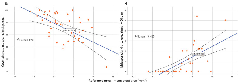
The average follow-up interval until OCT was 1580 ± 1260 days. Despite aorto-ostial stent protrusions in 40% of patients, optimal image quality was achieved in 93.3% of cases. There were higher rates of malapposed (11.4 ± 16.6 vs. 13.1 ± 8.3 vs. 0.3 ± 0.5%; < 0.001) and lower rates of covered struts (81.7 ± 16.8 vs. 83.7 ± 9.2 vs. 92.4 ± 6.8%; = 0.041) observed for the LM and POC segment compared to the dMB. Significantly malapposed stent struts (>400 μm) were less likely to be covered at follow-up, than struts with a measured strut to vessel wall distance of <400 μm (15.4 ± 21.6 vs. 24.8 ± 23.9%; = 0.011). Neoatherosclerosis was observed in 5 (33.3%) and restenotic neointimal hyperplasia (NIH) in 2 (13.3%) patients, requiring PCI in 33.3% of patients.

CONCLUSIONS

Long-term OCT examination of DES implanted during primary PCI for culprit ULM lesions demonstrated high rates of incomplete strut coverage, late malapposition, and high subclinical DES failure rates. These negative OCT results highlight the need for image optimization strategies during primary PCI to improve DES-related long-term outcomes.

摘要

背景

由于药物洗脱支架(DES)设计的进步、PCI技术的改进以及血管内成像的频繁使用,择期无保护左主干(ULM)经皮冠状动脉介入治疗(PCI)的长期死亡率与外科血运重建相当。然而,ULM罪犯病变的急诊PCI仍然与高院内死亡率和不良的长期预后相关,包括DES再狭窄和支架血栓形成(ST)。本分析旨在使用高分辨率光学相干断层扫描(OCT)成像检查在原发性PCI期间植入ULM的DES的长期预后和愈合情况。

方法

本分析纳入了来自一个高容量中心的15例连续接受ULM原发性PCI长期OCT随访的患者。在首次原发性PCI期间,所有受试者均接受了血管造影引导下的DES植入,除1例有非靶PCI病变的受试者外,所有受试者的随访均顺利。主要终点是长期随访时覆盖、未覆盖和贴壁不良的支架支柱百分比。次要终点包括定量和定性OCT测量。对于左主干分叉,在所有病例中对三个不同节段:左主干(LM)、汇合区(POC)和远端主支(dMB)进行了单独分析。

结果

直到进行OCT的平均随访间隔为1580±1260天。尽管40%的患者存在主动脉开口处支架突出,但93.3%的病例获得了最佳图像质量。与dMB相比,LM和POC节段观察到贴壁不良率更高(11.4±16.6 vs.13.1±8.3 vs.0.3±0.5%;P<0.001),覆盖支柱率更低(81.7±16.8 vs.83.7±9.2 vs.92.4±6.8%;P = 0.041)。与测量的支柱到血管壁距离<400μm的支柱相比,显著贴壁不良的支架支柱(>400μm)在随访时被覆盖的可能性更小(15.4±21.6 vs.24.8±23.9%;P = 0.011)。5例(33.3%)患者观察到新生动脉粥样硬化,2例(13.3%)患者观察到再狭窄性新生内膜增生(NIH),33.3%的患者需要进行PCI。

结论

对罪犯ULM病变进行原发性PCI期间植入的DES进行长期OCT检查显示,支架支柱覆盖不完全、晚期贴壁不良和亚临床DES失败率高。这些负面的OCT结果突出了在原发性PCI期间需要图像优化策略以改善DES相关的长期预后。

https://cdn.ncbi.nlm.nih.gov/pmc/blobs/63f0/11683725/3d6dd72e3d65/2153-8174-25-12-445-g6.jpg
https://cdn.ncbi.nlm.nih.gov/pmc/blobs/63f0/11683725/7aecda4b7079/2153-8174-25-12-445-g1.jpg
https://cdn.ncbi.nlm.nih.gov/pmc/blobs/63f0/11683725/a787ea18df8e/2153-8174-25-12-445-g2.jpg
https://cdn.ncbi.nlm.nih.gov/pmc/blobs/63f0/11683725/860af7ffcd46/2153-8174-25-12-445-g3.jpg
https://cdn.ncbi.nlm.nih.gov/pmc/blobs/63f0/11683725/95ca2593592e/2153-8174-25-12-445-g4.jpg
https://cdn.ncbi.nlm.nih.gov/pmc/blobs/63f0/11683725/19463c970144/2153-8174-25-12-445-g5.jpg
https://cdn.ncbi.nlm.nih.gov/pmc/blobs/63f0/11683725/3d6dd72e3d65/2153-8174-25-12-445-g6.jpg

相似文献

1
Long-term Follow-up Optical Coherence Tomography Assessment of Primary Percutaneous Coronary Intervention for Unprotected Left Main.无保护左主干原发性经皮冠状动脉介入治疗的长期随访光学相干断层扫描评估
Rev Cardiovasc Med. 2024 Dec 19;25(12):445. doi: 10.31083/j.rcm2512445. eCollection 2024 Dec.
2
Serial assessment of vessel interactions after drug-eluting stent implantation in unprotected distal left main coronary artery disease using frequency-domain optical coherence tomography.应用频域光学相干断层成像术对无保护的左主干冠状动脉远端病变行药物洗脱支架置入后血管相互作用的连续评估。
JACC Cardiovasc Interv. 2013 Oct;6(10):1035-45. doi: 10.1016/j.jcin.2013.05.015.
3
Incomplete stent apposition and delayed tissue coverage are more frequent in drug-eluting stents implanted during primary percutaneous coronary intervention for ST-segment elevation myocardial infarction than in drug-eluting stents implanted for stable/unstable angina: insights from optical coherence tomography.在急性 ST 段抬高型心肌梗死患者行经皮冠状动脉介入治疗时植入药物洗脱支架时,不完全贴壁和延迟组织覆盖比在稳定/不稳定型心绞痛患者中植入药物洗脱支架时更常见:光学相干断层扫描的见解。
JACC Cardiovasc Interv. 2009 May;2(5):445-52. doi: 10.1016/j.jcin.2009.01.012.
4
Optical coherence tomography analysis of the stent strut and prediction of resolved strut malapposition at 3 months after 2nd-generation drug-eluting stent implantation.第二代药物洗脱支架植入术后3个月时支架小梁的光学相干断层扫描分析及支架小梁贴壁不良消退的预测
Heart Vessels. 2016 Aug;31(8):1247-56. doi: 10.1007/s00380-015-0737-2. Epub 2015 Sep 3.
5
Difference in vascular response between sirolimus-eluting- and everolimus-eluting stents in ostial left circumflex artery after unprotected left main as observed by optical coherence tomography.光学相干断层扫描观察无保护左主干病变后左旋支开口处西罗莫司洗脱支架与依维莫司洗脱支架血管反应的差异
Int J Cardiol. 2017 Mar 1;230:284-292. doi: 10.1016/j.ijcard.2016.12.122. Epub 2016 Dec 23.
6
Three- and 6-month optical coherence tomographic surveillance following percutaneous coronary intervention with the Angiolite® drug-eluting stent: The ANCHOR study.使用Angiolite®药物洗脱支架经皮冠状动脉介入治疗后3个月和6个月的光学相干断层扫描监测:ANCHOR研究。
Catheter Cardiovasc Interv. 2018 Feb 15;91(3):435-443. doi: 10.1002/ccd.27189. Epub 2017 Jul 14.
7
Optical coherence tomography-based evaluation of malapposed strut coverage after drug-eluting stent implantation.基于光学相干断层扫描的药物洗脱支架置入后贴壁不良支架覆盖率评估。
Int J Cardiovasc Imaging. 2012 Dec;28(8):1887-94. doi: 10.1007/s10554-012-0039-z. Epub 2012 Mar 25.
8
Early vascular healing after neXt-generation drug-eluting stent implantation in Patients with non-ST elevation acute Coronary syndrome: a randomized optical coherence Tomography imaging study (EXPECT).非ST段抬高型急性冠状动脉综合征患者植入新一代药物洗脱支架后的早期血管愈合:一项随机光学相干断层扫描成像研究(EXPECT)
Int J Cardiovasc Imaging. 2024 Oct 12. doi: 10.1007/s10554-024-03258-w.
9
Optical Coherence Tomography Guided Percutaneous Coronary Intervention With Nobori Stent Implantation in Patients With Non-ST-Segment-Elevation Myocardial Infarction (OCTACS) Trial: Difference in Strut Coverage and Dynamic Malapposition Patterns at 6 Months.光学相干断层扫描指导下非ST段抬高型心肌梗死患者使用Nobori支架植入的经皮冠状动脉介入治疗(OCTACS)试验:6个月时支架小梁覆盖率及动态贴壁不良模式的差异
Circ Cardiovasc Interv. 2015 Aug;8(8):e002446. doi: 10.1161/CIRCINTERVENTIONS.114.002446.
10
The Cone Flare Crush Modified-T (CFCT) stenting technique for coronary artery bifurcation lesions.用于冠状动脉分叉病变的圆锥扩口挤压改良-T(CFCT)支架置入技术。
Int J Cardiol Heart Vasc. 2020 Sep 25;30:100643. doi: 10.1016/j.ijcha.2020.100643. eCollection 2020 Oct.

引用本文的文献

1
OCT for Optimizing Long-Term Clinical Results in Left Main PCI-Dream or Reality? Results from a Single-Center High-Volume Registry.光学相干断层扫描用于优化左主干经皮冠状动脉介入治疗的长期临床结果——梦想还是现实?来自单中心高容量注册研究的结果
J Clin Med. 2025 Aug 18;14(16):5824. doi: 10.3390/jcm14165824.

本文引用的文献

1
Percutaneous coronary intervention for bifurcation coronary lesions using optimised angiographic guidance: the 18th consensus document from the European Bifurcation Club.经皮冠状动脉介入治疗分叉病变的优化血管造影指导:来自欧洲分叉俱乐部的第 18 个共识文件。
EuroIntervention. 2024 Aug 5;20(15):e915-e926. doi: 10.4244/EIJ-D-24-00160.
2
Unintended Deformation of Stents During Bifurcation PCI: An OCTOBER Trial Substudy.支架在分叉 PCI 过程中的非计划性变形:OCTOBER 试验的子研究。
JACC Cardiovasc Interv. 2024 May 13;17(9):1106-1115. doi: 10.1016/j.jcin.2024.03.013.
3
OCT or Angiography Guidance for PCI in Complex Bifurcation Lesions.
OCT 或血管造影指导复杂分叉病变的 PCI
N Engl J Med. 2023 Oct 19;389(16):1477-1487. doi: 10.1056/NEJMoa2307770. Epub 2023 Aug 27.
4
2022 Joint ESC/EACTS review of the 2018 guideline recommendations on the revascularization of left main coronary artery disease in patients at low surgical risk and anatomy suitable for PCI or CABG.2022 年 ESC/EACTS 联合对 2018 年指南建议的回顾:低手术风险且适合 PCI 或 CABG 解剖结构的左主干冠状动脉疾病患者的血运重建。
Eur J Cardiothorac Surg. 2023 Aug 1;64(2). doi: 10.1093/ejcts/ezad286.
5
Management of failed stenting of the unprotected left main coronary artery.无保护左主干冠状动脉支架置入失败的处理。
Catheter Cardiovasc Interv. 2023 May;101(6):1001-1013. doi: 10.1002/ccd.30636. Epub 2023 Apr 1.
6
Optical Coherence Tomography to Assess Proximal Side Optimization Technique in Crush Stenting.光学相干断层扫描评估挤压式支架置入术中近端血管优化技术
Front Cardiovasc Med. 2022 Mar 15;9:861129. doi: 10.3389/fcvm.2022.861129. eCollection 2022.
7
2021 ACC/AHA/SCAI Guideline for Coronary Artery Revascularization: Executive Summary: A Report of the American College of Cardiology/American Heart Association Joint Committee on Clinical Practice Guidelines.2021 年 ACC/AHA/SCAI 冠状动脉血运重建指南:执行摘要:美国心脏病学会/美国心脏协会联合临床实践指南委员会的报告。
J Am Coll Cardiol. 2022 Jan 18;79(2):197-215. doi: 10.1016/j.jacc.2021.09.005. Epub 2021 Dec 9.
8
Clinical Implications of Poststent Optical Coherence Tomographic Findings: Severe Malapposition and Cardiac Events.支架置入术后光学相干断层扫描结果的临床意义:严重贴壁不良与心脏事件
JACC Cardiovasc Imaging. 2022 Jan;15(1):126-137. doi: 10.1016/j.jcmg.2021.03.008. Epub 2021 May 19.
9
Long-Term Clinical Outcomes of Unprotected Left Main Percutaneous Coronary Intervention: A Large Single-Centre Experience.无保护左主干经皮冠状动脉介入治疗的长期临床结果:一项大型单中心经验。
J Interv Cardiol. 2021 Jan 12;2021:8829686. doi: 10.1155/2021/8829686. eCollection 2021.
10
Optical coherence tomography to guide percutaneous coronary intervention of the left main coronary artery: the LEMON study.光学相干断层成像指导左主干冠状动脉经皮冠状动脉介入治疗:LEMON 研究。
EuroIntervention. 2021 Jun 11;17(2):e124-e131. doi: 10.4244/EIJ-D-20-01121.