Kim Seoyoung, Lee Young-Kwan, Lee Wang-Jun, Jong Moon Hyoun, Lee Sanghun
Eutilex Co. Ltd., Geumcheon-gu, Seoul, Republic of Korea.
Excelsisbio Inc., Goyang-si, Gyeonggi-do, Republic of Korea.
Integr Cancer Ther. 2025 Jan-Dec;24:15347354241308220. doi: 10.1177/15347354241308220.
Over the last decade, the anticancer effects of Stokes (RVS) have been reported in various preclinical or clinical studies. However, the effects of RVS on immuno-oncology, especially on the functional properties of T cells and their phenotypes, remain unclear. Here, we planned to investigate the impact of RVS on immuno-oncology, specifically focusing on its effects on T cells.
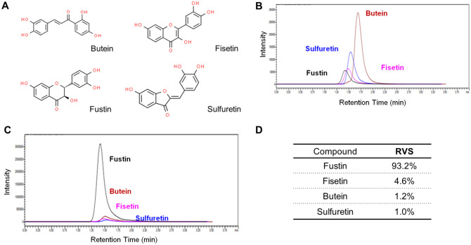
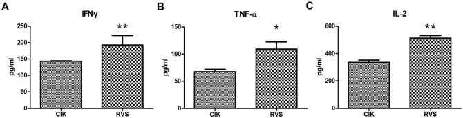
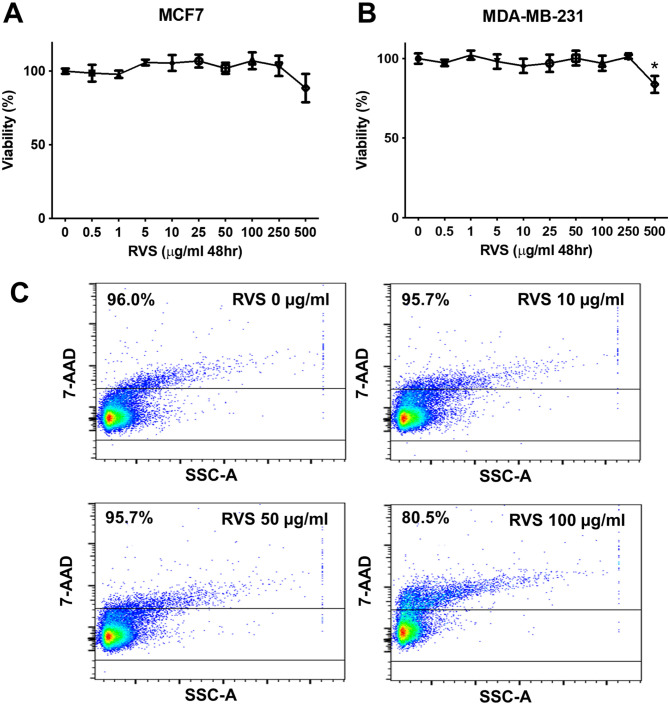
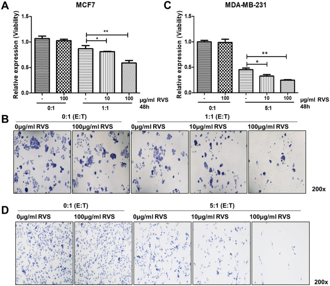
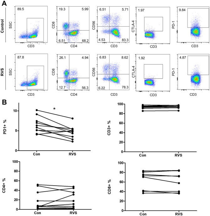
Peripheral blood mononuclear cells (PBMCs) from breast cancer patients were isolated to obtain cytokine-induced killer cell populations with >85% CD3+ T cells. The anticancer activity of these T cells was evaluated by introducing red fluorescent protein (RFP) into HLA-A02:01 type-matched breast cancer cell lines (MCF7 and MDA-MB-231) and analyzing the results using flow cytometry. The effect of RVS extracts on T cell phenotype was assessed using markers such as CTLA-4 and PD-1, as well as mRNA expression levels of key genes (IFN-γ, TNF-α, and IL-2).
RVS treatment significantly enhanced the anticancer activity of T cells against breast cancer cells. Specifically, T cells treated with 100 µg/mL of RVS showed a 20.6% increase in cytotoxicity against MCF-7 cells and a 36.2% increase against MDA-MB231 cells compared to the control. Additionally, RVS treatment led to a significant reduction in PD-1 expression on T cells.
Our findings demonstrate that RVS treatment enhances T cell function against breast cancer cells by reducing PD-1 expression. These results suggest that components of RVS may serve as potential candidates for restoring exhausted T cells in cancer therapy.
在过去十年中,多项临床前或临床研究报道了斯托克斯(RVS)的抗癌作用。然而,RVS对免疫肿瘤学的影响,尤其是对T细胞功能特性及其表型的影响仍不清楚。在此,我们计划研究RVS对免疫肿瘤学的影响,特别关注其对T细胞的作用。
分离乳腺癌患者的外周血单个核细胞(PBMC),以获得CD3+T细胞比例>85%的细胞因子诱导杀伤细胞群体。通过将红色荧光蛋白(RFP)导入HLA-A02:01型匹配的乳腺癌细胞系(MCF7和MDA-MB-231)并使用流式细胞术分析结果,评估这些T细胞的抗癌活性。使用CTLA-4和PD-1等标志物以及关键基因(IFN-γ、TNF-α和IL-2)的mRNA表达水平评估RVS提取物对T细胞表型的影响。
RVS处理显著增强了T细胞对乳腺癌细胞的抗癌活性。具体而言,与对照组相比,用100μg/mL RVS处理的T细胞对MCF-7细胞的细胞毒性增加了20.6%,对MDA-MB231细胞的细胞毒性增加了36.2%。此外,RVS处理导致T细胞上PD-1表达显著降低。
我们的研究结果表明,RVS处理通过降低PD-1表达增强了T细胞对乳腺癌细胞的功能。这些结果表明,RVS的成分可能是癌症治疗中恢复耗竭T细胞的潜在候选物。