• 文献检索
  • 文档翻译
  • 深度研究
  • 学术资讯
  • Suppr Zotero 插件Zotero 插件
  • 邀请有礼
  • 套餐&价格
  • 历史记录
应用&插件
Suppr Zotero 插件Zotero 插件浏览器插件Mac 客户端Windows 客户端微信小程序
定价
高级版会员购买积分包购买API积分包
服务
文献检索文档翻译深度研究API 文档MCP 服务
关于我们
关于 Suppr公司介绍联系我们用户协议隐私条款
关注我们

Suppr 超能文献

核心技术专利:CN118964589B侵权必究
粤ICP备2023148730 号-1Suppr @ 2026

文献检索

告别复杂PubMed语法,用中文像聊天一样搜索,搜遍4000万医学文献。AI智能推荐,让科研检索更轻松。

立即免费搜索

文件翻译

保留排版,准确专业,支持PDF/Word/PPT等文件格式,支持 12+语言互译。

免费翻译文档

深度研究

AI帮你快速写综述,25分钟生成高质量综述,智能提取关键信息,辅助科研写作。

立即免费体验

视网膜疾病早期检测的探索:基于3D循环生成对抗网络的光学相干断层扫描到共聚焦显微镜的转换

The quest for early detection of retinal disease: 3D CycleGAN-based translation of optical coherence tomography into confocal microscopy.

作者信息

Tian Xin, Anantrasirichai Nantheera, Nicholson Lindsay, Achim Alin

机构信息

Visual Information Laboratory, University of Bristol, Bristol, UK.

Autoimmune Inflammation Research, University of Bristol, Bristol, UK.

出版信息

Biol Imaging. 2024 Dec 16;4:e15. doi: 10.1017/S2633903X24000163. eCollection 2024.

DOI:10.1017/S2633903X24000163
PMID:39776613
原文链接:https://pmc.ncbi.nlm.nih.gov/articles/PMC11704141/
Abstract

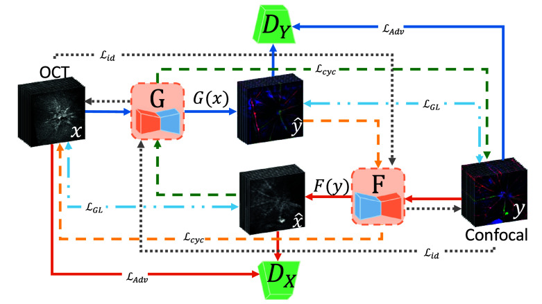
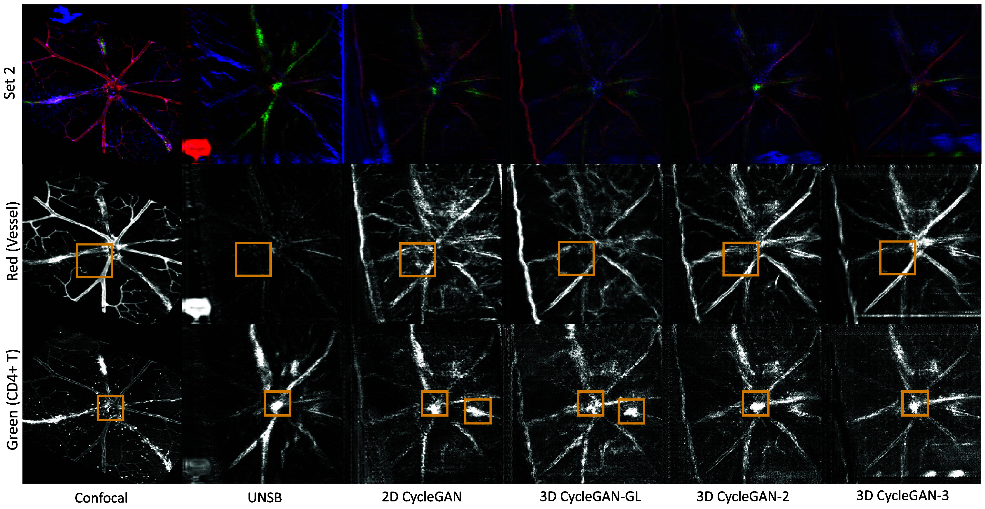
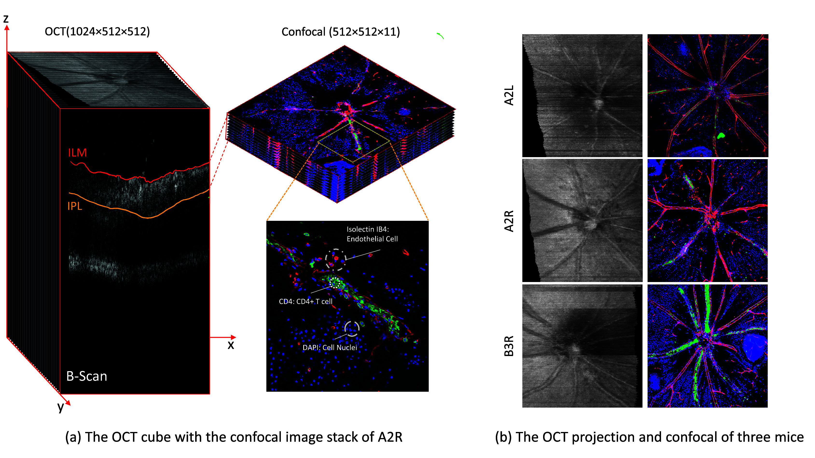
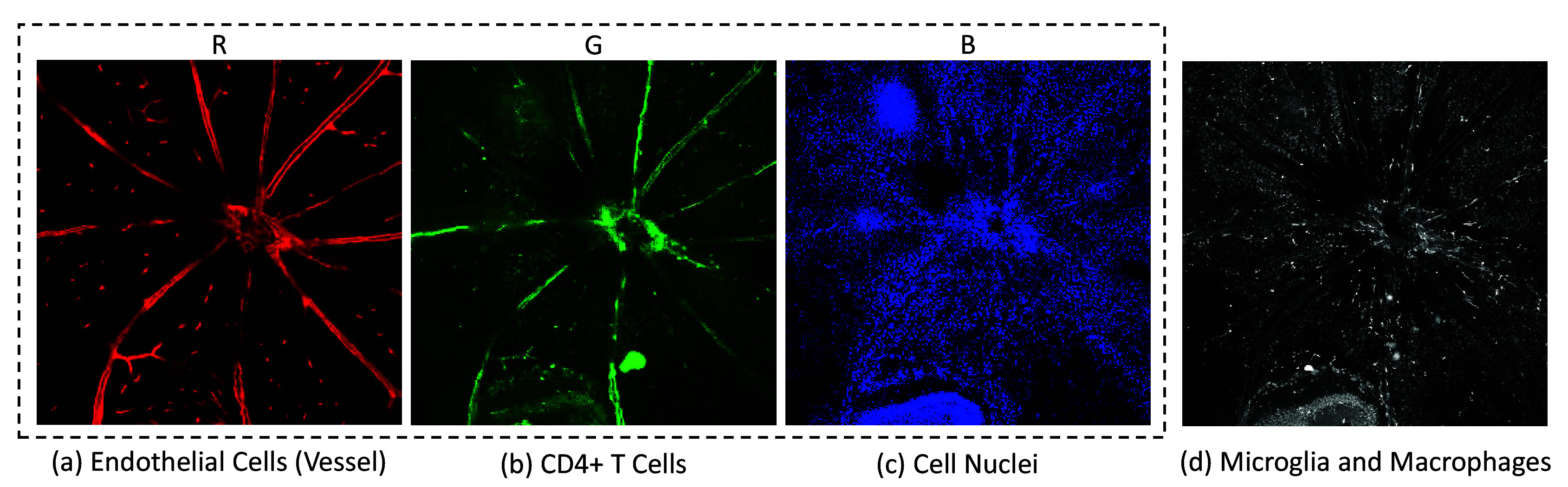
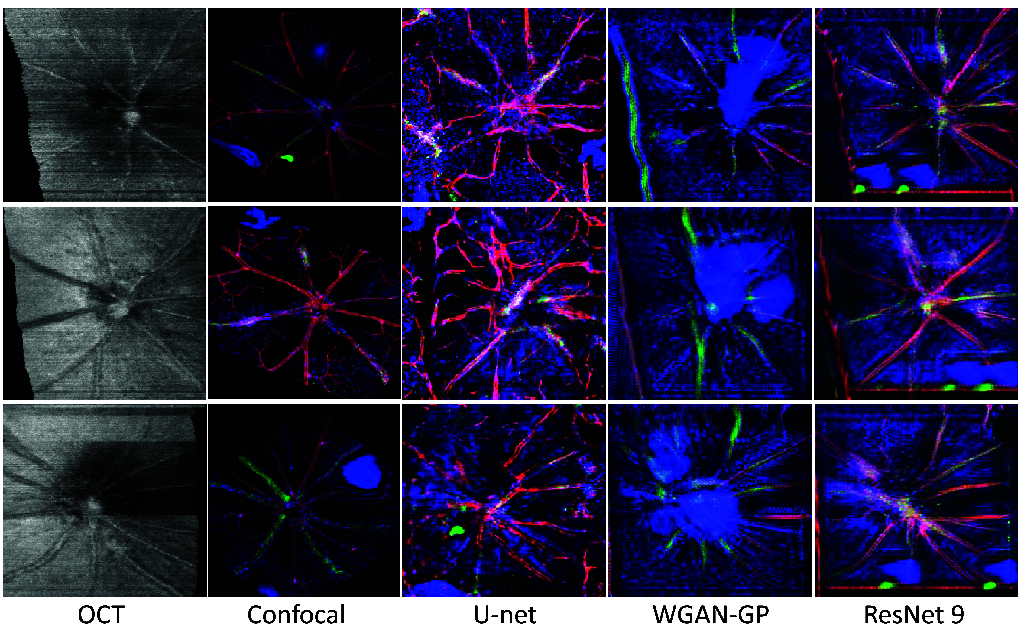
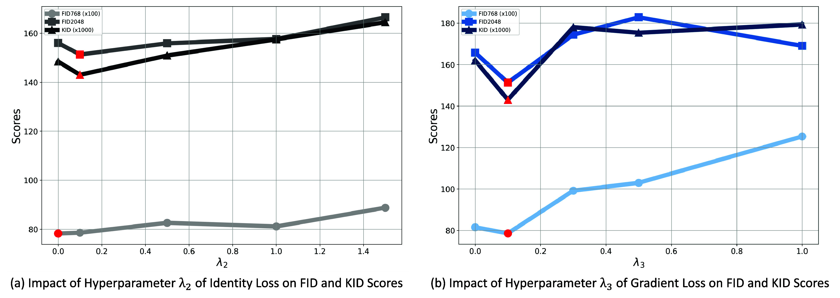
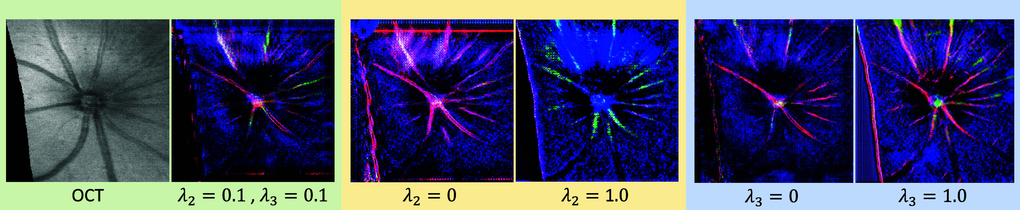
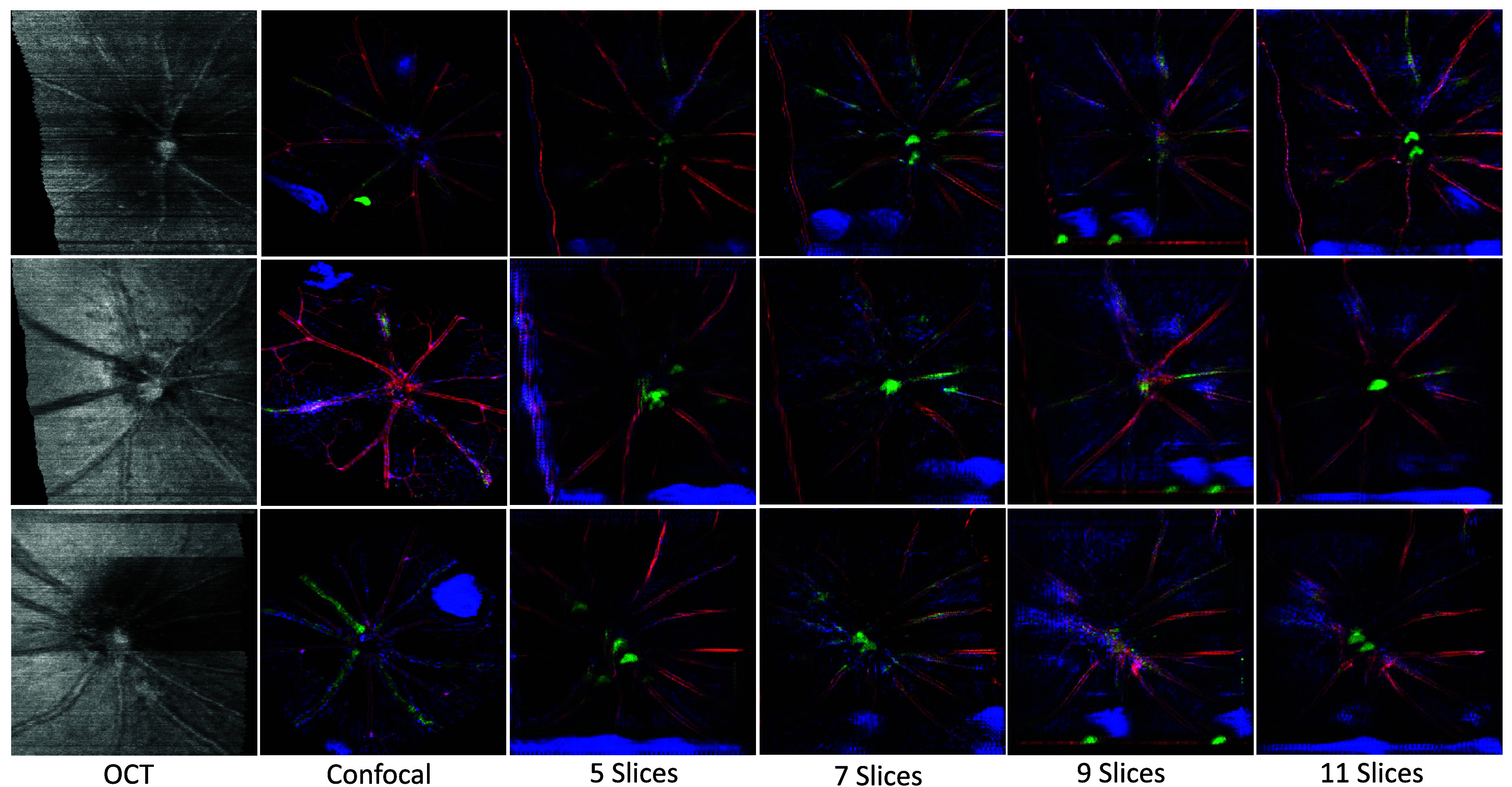
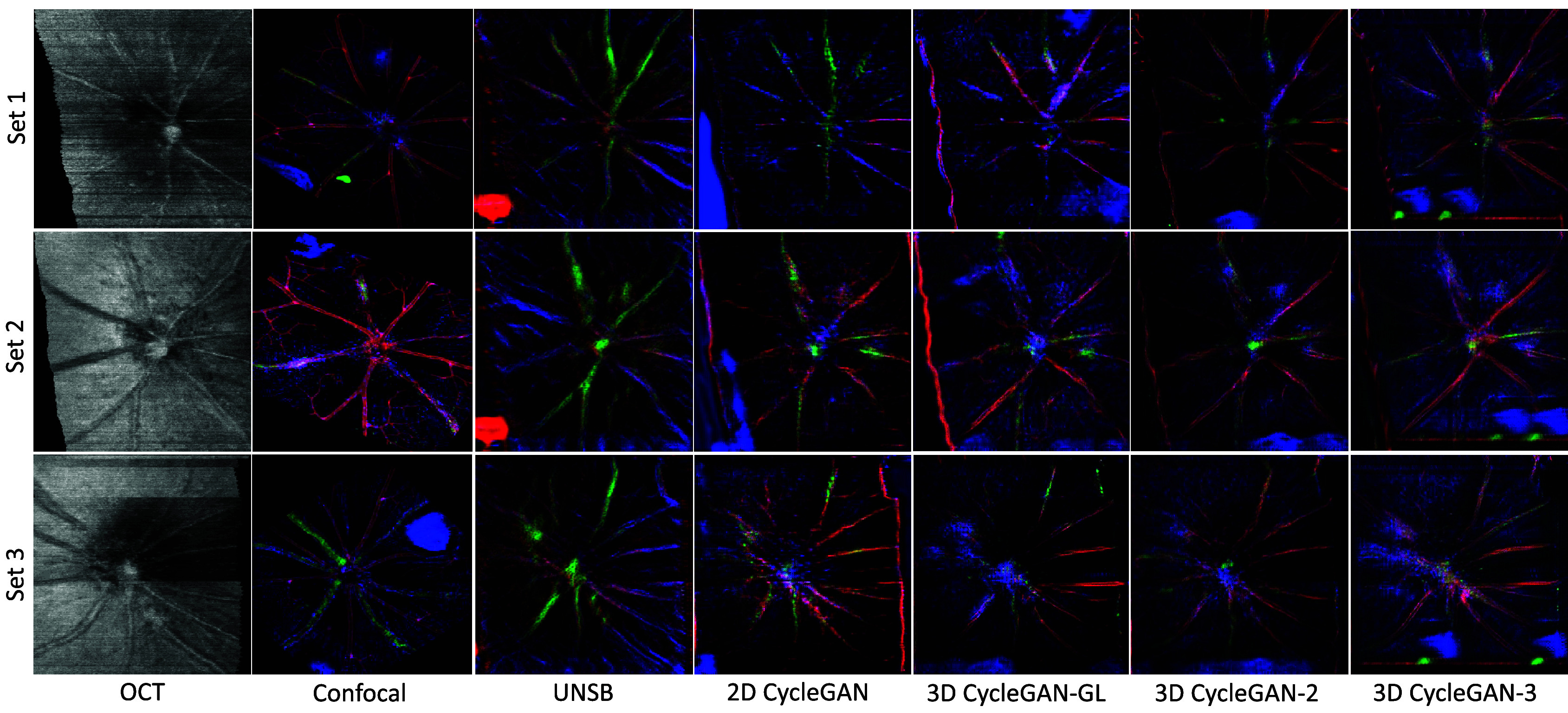
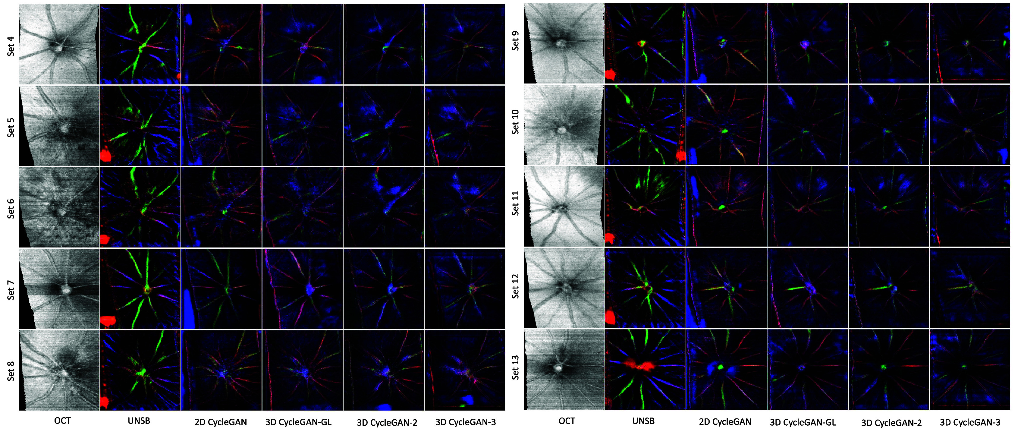
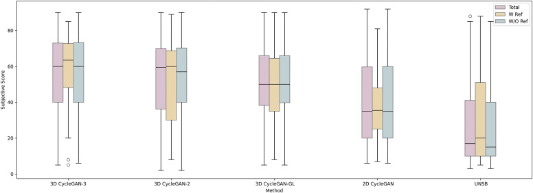
Optical coherence tomography (OCT) and confocal microscopy are pivotal in retinal imaging, offering distinct advantages and limitations. OCT offers rapid, noninvasive imaging but can suffer from clarity issues and motion artifacts, while confocal microscopy, providing high-resolution, cellular-detailed color images, is invasive and raises ethical concerns. To bridge the benefits of both modalities, we propose a novel framework based on unsupervised 3D CycleGAN for translating unpaired OCT to confocal microscopy images. This marks the first attempt to exploit the inherent 3D information of OCT and translate it into the rich, detailed color domain of confocal microscopy. We also introduce a unique dataset, OCT2Confocal, comprising mouse OCT and confocal retinal images, facilitating the development of and establishing a benchmark for cross-modal image translation research. Our model has been evaluated both quantitatively and qualitatively, achieving Fréchet inception distance (FID) scores of 0.766 and Kernel Inception Distance (KID) scores as low as 0.153, and leading subjective mean opinion scores (MOS). Our model demonstrated superior image fidelity and quality with limited data over existing methods. Our approach effectively synthesizes color information from 3D confocal images, closely approximating target outcomes and suggesting enhanced potential for diagnostic and monitoring applications in ophthalmology.

摘要

光学相干断层扫描(OCT)和共聚焦显微镜在视网膜成像中起着关键作用,各有其独特的优势和局限性。OCT提供快速、无创成像,但可能存在清晰度问题和运动伪影,而共聚焦显微镜虽能提供高分辨率、细胞细节丰富的彩色图像,却是侵入性的,还引发伦理问题。为融合这两种模式的优势,我们提出了一种基于无监督3D CycleGAN的新颖框架,用于将未配对的OCT图像转换为共聚焦显微镜图像。这是首次尝试利用OCT的固有3D信息并将其转换为共聚焦显微镜丰富、详细的彩色领域。我们还引入了一个独特的数据集OCT2Confocal,它由小鼠的OCT和共聚焦视网膜图像组成,有助于跨模态图像转换研究的发展并建立基准。我们的模型已进行了定量和定性评估,实现了0.766的弗雷歇 inception 距离(FID)分数以及低至0.153的核 inception 距离(KID)分数,并领先于主观平均意见分数(MOS)。与现有方法相比,我们的模型在有限数据下展示出卓越的图像保真度和质量。我们的方法有效地从3D共聚焦图像中合成颜色信息,非常接近目标结果,并显示出在眼科诊断和监测应用中增强的潜力。

https://cdn.ncbi.nlm.nih.gov/pmc/blobs/c050/11704141/b87146f27500/S2633903X24000163_fig11.jpg
https://cdn.ncbi.nlm.nih.gov/pmc/blobs/c050/11704141/3e4b9ec0d10f/S2633903X24000163_fig1.jpg
https://cdn.ncbi.nlm.nih.gov/pmc/blobs/c050/11704141/b68f4381993c/S2633903X24000163_fig2.jpg
https://cdn.ncbi.nlm.nih.gov/pmc/blobs/c050/11704141/ac62473f9602/S2633903X24000163_fig3.jpg
https://cdn.ncbi.nlm.nih.gov/pmc/blobs/c050/11704141/86fa65c9d18c/S2633903X24000163_fig4.jpg
https://cdn.ncbi.nlm.nih.gov/pmc/blobs/c050/11704141/6bc78a97fe68/S2633903X24000163_fig5.jpg
https://cdn.ncbi.nlm.nih.gov/pmc/blobs/c050/11704141/305f00006b5c/S2633903X24000163_fig6.jpg
https://cdn.ncbi.nlm.nih.gov/pmc/blobs/c050/11704141/e8118c9451b8/S2633903X24000163_fig7.jpg
https://cdn.ncbi.nlm.nih.gov/pmc/blobs/c050/11704141/7361ac631708/S2633903X24000163_fig8.jpg
https://cdn.ncbi.nlm.nih.gov/pmc/blobs/c050/11704141/b133ea136111/S2633903X24000163_fig9.jpg
https://cdn.ncbi.nlm.nih.gov/pmc/blobs/c050/11704141/84d933b76052/S2633903X24000163_fig10.jpg
https://cdn.ncbi.nlm.nih.gov/pmc/blobs/c050/11704141/b87146f27500/S2633903X24000163_fig11.jpg

相似文献

1
The quest for early detection of retinal disease: 3D CycleGAN-based translation of optical coherence tomography into confocal microscopy.视网膜疾病早期检测的探索:基于3D循环生成对抗网络的光学相干断层扫描到共聚焦显微镜的转换
Biol Imaging. 2024 Dec 16;4:e15. doi: 10.1017/S2633903X24000163. eCollection 2024.
2
Mirau-based line-field confocal optical coherence tomography for three-dimensional high-resolution skin imaging.基于 Mirau 的线场共焦光学相干层析成像术用于三维高分辨率皮肤成像。
J Biomed Opt. 2022 Aug;27(8). doi: 10.1117/1.JBO.27.8.086002.
3
A Method based on Evolutionary Algorithms and Channel Attention Mechanism to Enhance Cycle Generative Adversarial Network Performance for Image Translation.基于进化算法和通道注意力机制的方法来提高用于图像翻译的循环生成对抗网络性能。
Int J Neural Syst. 2023 May;33(5):2350026. doi: 10.1142/S0129065723500260. Epub 2023 Apr 5.
4
Generalized 3D registration algorithm for enhancing retinal optical coherence tomography images.用于增强视网膜光学相干断层扫描图像的广义三维配准算法。
J Biomed Opt. 2024 Jun;29(6):066002. doi: 10.1117/1.JBO.29.6.066002. Epub 2024 May 14.
5
Noise-imitation learning: unpaired speckle noise reduction for optical coherence tomography.噪声模拟学习:用于光学相干断层扫描的非配对散斑噪声减少。
Phys Med Biol. 2024 Sep 3;69(18). doi: 10.1088/1361-6560/ad708c.
6
Generative artificial intelligence to produce high-fidelity blastocyst-stage embryo images.生成式人工智能生成高保真囊胚期胚胎图像。
Hum Reprod. 2024 Jun 3;39(6):1197-1207. doi: 10.1093/humrep/deae064.
7
Three-dimensional retinal imaging with high-speed ultrahigh-resolution optical coherence tomography.采用高速超高分辨率光学相干断层扫描技术的三维视网膜成像
Ophthalmology. 2005 Oct;112(10):1734-46. doi: 10.1016/j.ophtha.2005.05.023.
8
Synthetic polarization-sensitive optical coherence tomography using contrastive unpaired translation.使用对比非配对翻译的合成偏振敏感光学相干断层扫描技术
Sci Rep. 2024 Dec 28;14(1):31366. doi: 10.1038/s41598-024-82839-0.
9
Triplet Cross-Fusion Learning for Unpaired Image Denoising in Optical Coherence Tomography.三重交叉融合学习在光学相干断层扫描中的未配对图像去噪。
IEEE Trans Med Imaging. 2022 Nov;41(11):3357-3372. doi: 10.1109/TMI.2022.3184529. Epub 2022 Oct 27.
10
In vivo three-dimensional high-resolution imaging of rodent retina with spectral-domain optical coherence tomography.利用谱域光学相干断层扫描技术对啮齿动物视网膜进行体内三维高分辨率成像。
Invest Ophthalmol Vis Sci. 2007 Apr;48(4):1808-14. doi: 10.1167/iovs.06-0815.

本文引用的文献

1
Multimodality Fusion Strategies in Eye Disease Diagnosis.多模态融合策略在眼病诊断中的应用
J Imaging Inform Med. 2024 Oct;37(5):2524-2558. doi: 10.1007/s10278-024-01105-x. Epub 2024 Apr 19.
2
Bright-field to fluorescence microscopy image translation for cell nuclei health quantification.用于细胞核健康定量的明场到荧光显微镜图像转换
Biol Imaging. 2023 Jun 15;3:e12. doi: 10.1017/S2633903X23000120. eCollection 2023.
3
Glaucoma: challenges and opportunities.青光眼:挑战与机遇
Clin Exp Optom. 2024 Mar;107(2):107-109. doi: 10.1080/08164622.2023.2300295. Epub 2024 Mar 11.
4
Translation of Color Fundus Photography into Fluorescein Angiography Using Deep Learning for Enhanced Diabetic Retinopathy Screening.利用深度学习将彩色眼底照片转换为荧光血管造影以增强糖尿病视网膜病变筛查
Ophthalmol Sci. 2023 Sep 15;3(4):100401. doi: 10.1016/j.xops.2023.100401. eCollection 2023 Dec.
5
Artificial intelligence in the diagnosis of glaucoma and neurodegenerative diseases.人工智能在青光眼和神经退行性疾病诊断中的应用。
Clin Exp Optom. 2024 Mar;107(2):130-146. doi: 10.1080/08164622.2023.2235346. Epub 2023 Sep 6.
6
OCT-based deep-learning models for the identification of retinal key signs.基于 OCT 的深度学习模型用于识别视网膜关键征象。
Sci Rep. 2023 Sep 5;13(1):14628. doi: 10.1038/s41598-023-41362-4.
7
A machine learning framework for the quantification of experimental uveitis in murine OCT.一种用于量化小鼠光学相干断层扫描(OCT)中实验性葡萄膜炎的机器学习框架。
Biomed Opt Express. 2023 Jun 16;14(7):3413-3432. doi: 10.1364/BOE.489271. eCollection 2023 Jul 1.
8
Medical image data augmentation: techniques, comparisons and interpretations.医学图像数据增强:技术、比较与解读
Artif Intell Rev. 2023 Mar 20:1-45. doi: 10.1007/s10462-023-10453-z.
9
Cross-modality image translation: CT image synthesis of MR brain images using multi generative network with perceptual supervision.跨模态图像翻译:使用具有感知监督的多生成网络对 MR 脑图像进行 CT 图像合成。
Comput Methods Programs Biomed. 2023 Jul;237:107571. doi: 10.1016/j.cmpb.2023.107571. Epub 2023 Apr 30.
10
Deep learning based unpaired image-to-image translation applications for medical physics: a systematic review.基于深度学习的医学物理非配对图像到图像转换应用:系统综述
Phys Med Biol. 2023 Feb 23;68(5). doi: 10.1088/1361-6560/acba74.