• 文献检索
  • 文档翻译
  • 深度研究
  • 学术资讯
  • Suppr Zotero 插件Zotero 插件
  • 邀请有礼
  • 套餐&价格
  • 历史记录
应用&插件
Suppr Zotero 插件Zotero 插件浏览器插件Mac 客户端Windows 客户端微信小程序
定价
高级版会员购买积分包购买API积分包
服务
文献检索文档翻译深度研究API 文档MCP 服务
关于我们
关于 Suppr公司介绍联系我们用户协议隐私条款
关注我们

Suppr 超能文献

核心技术专利:CN118964589B侵权必究
粤ICP备2023148730 号-1Suppr @ 2026

文献检索

告别复杂PubMed语法,用中文像聊天一样搜索,搜遍4000万医学文献。AI智能推荐,让科研检索更轻松。

立即免费搜索

文件翻译

保留排版,准确专业,支持PDF/Word/PPT等文件格式,支持 12+语言互译。

免费翻译文档

深度研究

AI帮你快速写综述,25分钟生成高质量综述,智能提取关键信息,辅助科研写作。

立即免费体验

超声造影在乳腺可疑病变鉴别诊断中的应用价值

The benefits of contrast-enhanced ultrasound in the differential diagnosis of suspicious breast lesions.

作者信息

Liang Runa, Lian Jun, Zhang Jinhui, Jing Jiayu, Bian Jinxia, Xu Jinzhi, He Xin, Yu Shanshan, Zhou Qi, Jiang Jue

机构信息

Department of Ultrasound, The Second Affiliated Hospital of Xi'an Jiaotong University, Xi'an, China.

Department of Ultrasound, Ankang Central Hospital, Ankang, China.

出版信息

Front Med (Lausanne). 2024 Dec 24;11:1511200. doi: 10.3389/fmed.2024.1511200. eCollection 2024.

DOI:10.3389/fmed.2024.1511200
PMID:39776839
原文链接:https://pmc.ncbi.nlm.nih.gov/articles/PMC11703730/
Abstract

BACKGROUND

Contrast-enhanced ultrasound (CEUS) shows potential for the differential diagnosis of breast lesions in general, but its effectiveness remains unclear for the differential diagnosis of lesions highly suspicious for breast cancers.

OBJECTIVE

This study aimed to evaluate the diagnostic value of CEUS in differentiating pathological subtypes of suspicious breast lesions defined as category 4 of US-BI-RADS.

METHODS

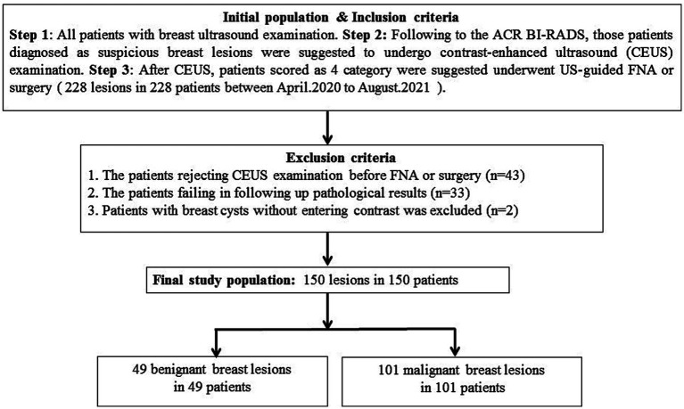
The dataset of 150 breast lesions was prospectively collected from 150 patients who underwent routine ultrasound and CEUS examination and were highly suspected of having breast cancers. All lesions were pathologically confirmed by US-guided needle biopsy and surgery. The qualitative features and the quantitative parameters of CEUS of these breast lesions were analyzed. The CEUS and biopsy examinations were performed after informed consent.

RESULTS

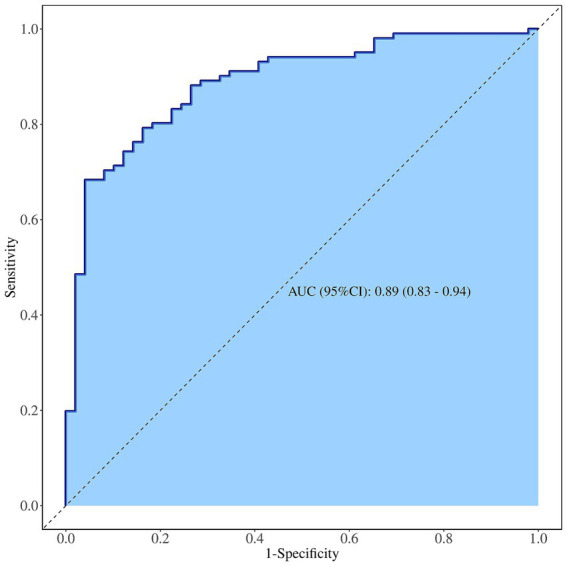
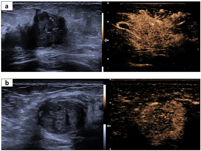
In the qualitative features, crab clam-like enhancement, the presence of more than two enhanced vessels within lesions, and surrounding enriched vessels inserting into lesions were able to differentiate atypical fibroadenomas (FIB) and mass-like non-puerperal mastitis (NPM) from invasive ductal carcinomas (IDC) and ductal carcinomas (DCIS) ( < 0.05). The enlarged scope, irregular shape, and perfusion deficiency were valuable to the differential diagnosis of FIB from the others ( < 0.05). In the four quantitative parameters of CEUS, only the peak intensity (IMAX) contributed to the differential diagnosis between malignant and benign tumors ( < 0.05, ROCAUC: 0.61, sensitivity: 60.4% and specificity: 65.9%, accuracy: 62.1%). However, IMAX did not show any difference in the paired comparison of IDC, DCIS, FIB, and NPM ( > 0.05). The logistic regression analysis results showed that heterogeneous perfusion, crab clam-like enhancement, and partial_ IMAX were independent risk factors for benign and malignant breast lesions . The area under a receiver operating characteristic of the integrated model was 0.89. In the diagnosis of benign and malignant pathological subtypes of breast lesions, independent risk factors and integrated models had no statistical significance in the diagnosis of IDC and DCISs, FIB, and NPM ( > 0.05).

CONCLUSION

Some qualitative risk features of CEUS can distinguish malignant breast lesions from NPM and atypical FIB with a high score of US-BI-RADS, aiding physicians to reduce the misdiagnosis of suspicious breast lesions in clinical practice.

摘要

背景

一般而言,超声造影(CEUS)在乳腺病变的鉴别诊断中显示出潜力,但其在高度怀疑为乳腺癌的病变鉴别诊断中的有效性仍不明确。

目的

本研究旨在评估CEUS在鉴别美国放射学会乳腺影像报告和数据系统(US-BI-RADS)4类可疑乳腺病变病理亚型中的诊断价值。

方法

前瞻性收集150例接受常规超声和CEUS检查且高度怀疑患有乳腺癌患者的150个乳腺病变数据集。所有病变均经超声引导下穿刺活检和手术病理证实。分析这些乳腺病变CEUS的定性特征和定量参数。在获得知情同意后进行CEUS和活检检查。

结果

在定性特征方面,蟹钳样增强、病灶内有两条以上增强血管以及周围有丰富血管穿入病灶能够将非典型纤维腺瘤(FIB)和肿块样非产褥期乳腺炎(NPM)与浸润性导管癌(IDC)和导管原位癌(DCIS)区分开来(P<0.05)。范围扩大、形态不规则和灌注不足对FIB与其他病变的鉴别诊断有价值(P<0.05)。在CEUS的四个定量参数中,只有峰值强度(IMAX)对良恶性肿瘤的鉴别诊断有贡献(P<0.05,受试者工作特征曲线下面积:0.61,灵敏度:60.4%,特异度:65.9%,准确度:62.1%)。然而,IMAX在IDC、DCIS、FIB和NPM的配对比较中未显示出任何差异(P>0.05)。逻辑回归分析结果显示,不均匀灌注、蟹钳样增强和部分IMAX是乳腺良恶性病变的独立危险因素。综合模型的受试者工作特征曲线下面积为0.89。在乳腺病变良恶性病理亚型的诊断中,独立危险因素和综合模型在IDC、DCIS、FIB和NPM的诊断中无统计学意义(P>0.05)。

结论

CEUS的一些定性风险特征可以将乳腺恶性病变与NPM和US-BI-RADS分类为4类的非典型FIB区分开来,有助于医生在临床实践中减少可疑乳腺病变的误诊。

https://cdn.ncbi.nlm.nih.gov/pmc/blobs/fc09/11703730/b45a735b2fe4/fmed-11-1511200-g004.jpg
https://cdn.ncbi.nlm.nih.gov/pmc/blobs/fc09/11703730/50a25abef1c8/fmed-11-1511200-g001.jpg
https://cdn.ncbi.nlm.nih.gov/pmc/blobs/fc09/11703730/baf5128ef33a/fmed-11-1511200-g002.jpg
https://cdn.ncbi.nlm.nih.gov/pmc/blobs/fc09/11703730/bd58ce986d36/fmed-11-1511200-g003.jpg
https://cdn.ncbi.nlm.nih.gov/pmc/blobs/fc09/11703730/b45a735b2fe4/fmed-11-1511200-g004.jpg

相似文献

1
The benefits of contrast-enhanced ultrasound in the differential diagnosis of suspicious breast lesions.超声造影在乳腺可疑病变鉴别诊断中的应用价值
Front Med (Lausanne). 2024 Dec 24;11:1511200. doi: 10.3389/fmed.2024.1511200. eCollection 2024.
2
The diagnostic value of contrast-enhanced ultrasonography in breast ductal abnormalities.增强超声在乳腺导管异常中的诊断价值。
Cancer Imaging. 2023 Mar 10;23(1):25. doi: 10.1186/s40644-023-00539-w.
3
Diagnostic test of conventional ultrasonography combined with contrast-enhanced ultrasound in the subcategorization of suspicious Breast Imaging-Reporting and Data System (BI-RADS) 4 breast lesions.常规超声联合超声造影对可疑乳腺影像报告和数据系统(BI-RADS)4类乳腺病变进行亚分类的诊断试验
Transl Cancer Res. 2025 Mar 30;14(3):2066-2077. doi: 10.21037/tcr-2025-485. Epub 2025 Mar 27.
4
The diagnostic value of contrast-enhanced ultrasound combined with clinicopathological features in microinvasive ductal carcinoma .超声造影联合临床病理特征在微浸润性导管癌中的诊断价值
Gland Surg. 2024 Nov 30;13(11):1894-1906. doi: 10.21037/gs-24-211. Epub 2024 Nov 26.
5
Breast non-mass-like lesions on contrast-enhanced ultrasonography: Feature analysis, breast image reporting and data system classification assessment.超声造影下乳腺非肿块样病变:特征分析、乳腺影像报告和数据系统分类评估
World J Clin Cases. 2020 Feb 26;8(4):700-712. doi: 10.12998/wjcc.v8.i4.700.
6
Application of contrast-enhanced ultrasound in the diagnosis of small breast lesions.超声造影在乳腺小病灶诊断中的应用
Clin Hemorheol Microcirc. 2018;70(3):291-300. doi: 10.3233/CH-170368.
7
Predictive value of contrast-enhanced ultrasonography and ultrasound elastography for management of BI-RADS category 4 nonpalpable breast masses.超声造影及超声弹性成像对 BI-RADS 分类 4 类触诊阴性乳腺肿块的预测价值。
Eur J Radiol. 2024 Apr;173:111391. doi: 10.1016/j.ejrad.2024.111391. Epub 2024 Feb 28.
8
Qualitative, quantitative and combination score systems in differential diagnosis of breast lesions by contrast-enhanced ultrasound.超声造影在乳腺病变鉴别诊断中的定性、定量及联合评分系统
Eur J Radiol. 2016 Jan;85(1):48-54. doi: 10.1016/j.ejrad.2015.10.017. Epub 2015 Oct 31.
9
Diagnostic accuracy of contrast-enhanced ultrasound synchronized with shear wave elastography in the differential diagnosis of benign and malignant breast lesions: a diagnostic test.超声造影与剪切波弹性成像同步在乳腺良恶性病变鉴别诊断中的诊断准确性:一项诊断试验
Gland Surg. 2023 Jan 1;12(1):54-66. doi: 10.21037/gs-22-684. Epub 2023 Jan 13.
10
A Prospective Multicenter Study on the Additive Value of Contrast-Enhanced Ultrasound for Biopsy Decision of Ultrasound BI-RADS 4 Breast Lesions.对比增强超声在超声 BI-RADS 4 类乳腺病变活检决策中附加价值的前瞻性多中心研究。
Ultrasound Med Biol. 2024 Aug;50(8):1224-1231. doi: 10.1016/j.ultrasmedbio.2024.04.010. Epub 2024 May 24.

本文引用的文献

1
Meta-analysis of contrast-enhanced ultrasound in differential diagnosis of breast adenosis and breast cancer.超声造影增强模式对乳腺腺病与乳腺癌的鉴别诊断价值的Meta 分析
J Clin Ultrasound. 2024 Nov-Dec;52(9):1402-1418. doi: 10.1002/jcu.23803. Epub 2024 Aug 29.
2
Global cancer statistics 2022: GLOBOCAN estimates of incidence and mortality worldwide for 36 cancers in 185 countries.2022 年全球癌症统计数据:全球 185 个国家和地区 36 种癌症的发病率和死亡率全球估计数。
CA Cancer J Clin. 2024 May-Jun;74(3):229-263. doi: 10.3322/caac.21834. Epub 2024 Apr 4.
3
Diagnostic performance of contrast-enhanced ultrasound combined with shear wave elastography in differentiating benign from malignant breast lesions: a systematic review and meta-analysis.
超声造影联合剪切波弹性成像在鉴别乳腺良恶性病变中的诊断性能:一项系统评价和荟萃分析
Gland Surg. 2023 Nov 24;12(11):1610-1623. doi: 10.21037/gs-23-333. Epub 2023 Oct 20.
4
A Nomogram Based on Conventional and Contrast-Enhanced Ultrasound for Pre-operative Prediction of Nipple-Areola Complex Involvement in Breast Cancer: A Prospective Study.基于常规超声和超声造影的术前预测乳腺癌乳头乳晕复合体受累的列线图:一项前瞻性研究。
Ultrasound Med Biol. 2023 Dec;49(12):2565-2572. doi: 10.1016/j.ultrasmedbio.2023.08.023. Epub 2023 Sep 24.
5
Diagnostic accuracy of contrast-enhanced ultrasound synchronized with shear wave elastography in the differential diagnosis of benign and malignant breast lesions: a diagnostic test.超声造影与剪切波弹性成像同步在乳腺良恶性病变鉴别诊断中的诊断准确性:一项诊断试验
Gland Surg. 2023 Jan 1;12(1):54-66. doi: 10.21037/gs-22-684. Epub 2023 Jan 13.
6
Quantification and Classification of Contrast Enhanced Ultrasound Breast Cancer Data: A Preliminary Study.超声造影增强乳腺癌数据的量化与分类:一项初步研究。
Diagnostics (Basel). 2022 Feb 6;12(2):425. doi: 10.3390/diagnostics12020425.
7
Performance of Contrast-Enhanced Ultrasound in Thyroid Nodules: Review of Current State and Future Perspectives.超声造影在甲状腺结节中的应用:现状与未来展望
Cancers (Basel). 2021 Oct 30;13(21):5469. doi: 10.3390/cancers13215469.
8
Real-life assessment of standardized contrast-enhanced ultrasound (CEUS) and CEUS algorithms (CEUS LI-RADS®/ESCULAP) in hepatic nodules in cirrhotic patients-a prospective multicenter study.肝硬化患者肝结节中标准化对比增强超声(CEUS)及 CEUS 算法(CEUS LI-RADS®/ESCULAP)的真实世界评估 - 一项前瞻性多中心研究。
Eur Radiol. 2021 Oct;31(10):7614-7625. doi: 10.1007/s00330-021-07872-3. Epub 2021 Apr 15.
9
Predictive risk factors for sentinel lymph node metastasis using preoperative contrast-enhanced ultrasound in early-stage breast cancer patients.早期乳腺癌患者术前使用超声造影预测前哨淋巴结转移的危险因素
Gland Surg. 2021 Feb;10(2):761-769. doi: 10.21037/gs-20-867.
10
Prospective evaluation of contrast-enhanced ultrasound of breast BI-RADS 3-5 lesions.乳腺影像报告和数据系统(BI-RADS)3 - 5类病变的超声造影前瞻性评估
BMC Med Imaging. 2020 Jun 17;20(1):66. doi: 10.1186/s12880-020-00467-2.