Reynard M, Riffenburgh R S, Minckler D S
Br J Ophthalmol. 1985 Mar;69(3):197-201. doi: 10.1136/bjo.69.3.197.
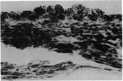
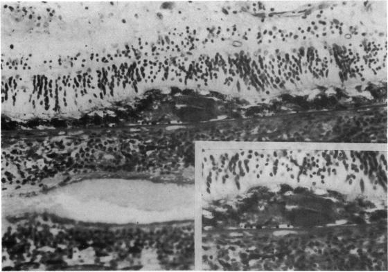
Fifty cases of sympathetic ophthalmia were examined histologically to determine the incidence and morphology of Dalén-Fuchs nodules. At least one well-defined Dalén-Fuchs nodule was identified in 18 (36%) of the eyes examined. Three types of lesions at the level of the retinal pigment epithelium were recognised. One type was found to consist of focal hyperplasia and aggregation of retinal pigment epithelial cells. A second type, classically referred to as Dalén-Fuchs nodules, consisted of epithelioid cells and lymphocytes covered by an intact dome of retinal pigment epithelium. The third type of lesion was characterised by degeneration of the overlying retinal pigment epithelium leading to disorganisation of the Dalén-Fuchs nodule and possible release of their contents into the subretinal space. Our study demonstrated that Dalén-Fuchs nodules in sympathetic ophthalmia vary in their morphological appearance as determined by light microscopy.
对50例交感性眼炎患者进行了组织学检查,以确定达伦-富克斯结节的发生率和形态。在所检查的眼睛中,18只(36%)至少发现了一个界限清楚的达伦-富克斯结节。在视网膜色素上皮水平识别出三种类型的病变。一种类型是视网膜色素上皮细胞的局灶性增生和聚集。第二种类型,经典地称为达伦-富克斯结节,由上皮样细胞和淋巴细胞组成,被完整的视网膜色素上皮穹顶覆盖。第三种类型的病变特征是上方视网膜色素上皮变性,导致达伦-富克斯结节结构紊乱,并可能使其内容物释放到视网膜下间隙。我们的研究表明,通过光学显微镜观察,交感性眼炎中的达伦-富克斯结节在形态外观上存在差异。