Gon Hidetoshi, Omiya Satoshi, Komatsu Shohei, Yamasaki Nobuaki, Murakami Sae, Fukushima Kenji, Urade Takeshi, Tsugawa Daisuke, Yanagimoto Hiroaki, Toyama Hirochika, Kido Masahiro, Fukumoto Takumi
Department of Surgery, Division of Hepato-Biliary-Pancreatic Surgery, Kobe University Graduate School of Medicine, Kobe, Hyogo, Japan.
Clinical and Translational Research Center, Kobe University Hospital, Kobe, Hyogo, Japan.
Langenbecks Arch Surg. 2025 Jan 11;410(1):34. doi: 10.1007/s00423-024-03602-7.
This study aimed to evaluate the efficacy of indocyanine green (ICG)-fluorescence imaging for the identification of hepatic boundaries during liver resection and its advantages in surgical outcomes over conventional methods.
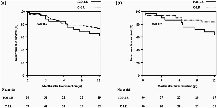
This prospective, exploratory, single-arm clinical trial included 47 patients with liver tumors who underwent liver resection using ICG-fluorescence imaging (ICG-LR) between 2019 and 2020. The primary outcome measure was the successful identification of hepatic boundaries during liver resection, from the perspective of both the hepatic surface and intrahepatic boundary, using ICG-fluorescence imaging. The secondary outcomes comprised surgical outcomes. Using propensity score matching (PSM), the surgical outcomes were subsequently compared between the ICG-LR group and patients who underwent conventional liver resection (C-LR, n = 100) between 2017 and 2018.
Hepatic boundaries were successfully identified in 28 patients (60%; 95% confidence interval, 45-72%), including 21 and 7 who underwent anatomical and non-anatomical liver resection, respectively. After PSM, 40 patients were included in each of the ICG-LR and C-LR groups. The surgical outcomes were similar between the groups. Subsequently, surgical outcomes were compared between the groups focusing on anatomical liver resection. After PSM, 21 patients were included in each group. The ICG-LR group had a lower rate of Clavien-Dindo grade ≥ IIIa complications (0% vs. 24%; P = 0.017), including ascites and bile leak, and a shorter hospital stay (12 vs. 14 days, P = 0.041) than the C-LR group did.
ICG-fluorescence imaging could be used to recognize hepatic boundaries during liver transection. Additionally, ICG-LR may be useful in preventing severe liver-associated complications.
This study is registered at the UMIN Clinical Trials Registry: UMIN0000180139 and Japan Registry of Clinical Trials: jRCT1051180070. The Registration Data Set is available at https://jrct.niph.go.jp/ .
本研究旨在评估吲哚菁绿(ICG)荧光成像在肝切除术中识别肝边界的有效性及其在手术结果方面相对于传统方法的优势。
这项前瞻性、探索性、单臂临床试验纳入了47例肝肿瘤患者,这些患者在2019年至2020年期间接受了使用ICG荧光成像(ICG-LR)的肝切除术。主要结局指标是从肝表面和肝内边界两个角度,使用ICG荧光成像在肝切除术中成功识别肝边界。次要结局包括手术结果。随后,使用倾向评分匹配(PSM)对ICG-LR组与2017年至2018年期间接受传统肝切除术(C-LR,n = 100)的患者的手术结果进行比较。
28例患者(60%;95%置信区间,45 - 72%)成功识别了肝边界,其中分别有21例和7例接受了解剖性和非解剖性肝切除术。PSM后,ICG-LR组和C-LR组各纳入40例患者。两组的手术结果相似。随后,重点比较了两组解剖性肝切除术的手术结果。PSM后,每组纳入21例患者。ICG-LR组Clavien-Dindo≥IIIa级并发症(包括腹水和胆漏)的发生率低于C-LR组(0%对24%;P = 0.017),住院时间也更短(12天对14天,P = 0.041)。
ICG荧光成像可用于在肝横断术中识别肝边界。此外,ICG-LR可能有助于预防严重的肝脏相关并发症。
本研究已在UMIN临床试验注册中心注册:UMIN0000180139,以及日本临床试验注册中心:jRCT1051180070。注册数据集可在https://jrct.niph.go.jp/获取。