Department of Surgery, Division of Hepato-Biliary-Pancreatic Surgery, Kobe University Graduate School of Medicine, Kobe, Japan
Department of Surgery, Division of Hepato-Biliary-Pancreatic Surgery, Kobe University Graduate School of Medicine, Kobe, Japan.
BMJ Open. 2019 Aug 18;9(8):e030233. doi: 10.1136/bmjopen-2019-030233.
In-vivo fluorescence imaging techniques using indocyanine green (ICG) to identify liver tumours and hepatic segment boundaries have been recently developed. The purpose of this study is to evaluate the efficacy of fusion ICG-fluorescence imaging for navigation during hepatectomy.
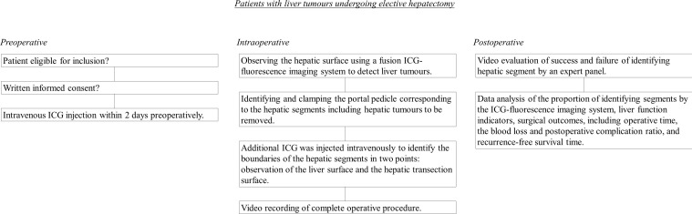
This will be an exploratory single-arm clinical trial; patients with liver tumours will undergo hepatectomy using the ICG-fluorescence imaging system. In total, 110 patients with liver tumours scheduled for elective hepatectomy will be included in this study. Preoperatively, ICG will be intravenously injected at a dose of 0.5 mg/kg body weight within 2 days. To detect liver tumours intraoperatively, the hepatic surface will be initially observed using the ICG-fluorescence imaging system. After identifying and clamping the portal pedicle corresponding to the hepatic segments, including the liver tumours to be resected, additional ICG will be injected intravenously at a dose of 0.5 mg/kg body weight to identify the boundaries of the hepatic segments. The primary outcome measure will be the success or failure of the ICG-fluorescence imaging system in identifying hepatic segments. The secondary outcomes will be the success or failure in identifying liver tumours, liver function indicators, operative time, blood loss, rate of postoperative complications and recurrence-free survival. The findings obtained through this study are expected to help to establish the utility of ICG-fluorescence imaging systems, and therefore contribute to prognostic outcome improvements in patients undergoing hepatectomy for various causes.
The protocol has been approved by the Kobe University Clinical Research Ethical Committee. The findings of this study will be disseminated widely through peer-reviewed publications and conference presentations.
UMIN000031054 and jRCT1051180070.
最近已经开发出了使用吲哚菁绿(ICG)进行体内荧光成像技术,以识别肝肿瘤和肝段边界。本研究的目的是评估融合 ICG-荧光成像在肝切除术中导航的效果。
这将是一项探索性的单臂临床试验;接受肝肿瘤切除术的患者将使用 ICG-荧光成像系统进行手术。总共将有 110 名计划接受择期肝切除术的肝肿瘤患者纳入本研究。术前,将在 2 天内以 0.5mg/kg 体重的剂量静脉注射 ICG。为了在术中检测肝肿瘤,将首先使用 ICG-荧光成像系统观察肝表面。在识别和夹闭包括待切除肝肿瘤的肝段相应的门脉蒂后,将以 0.5mg/kg 体重的剂量静脉注射额外的 ICG,以识别肝段的边界。主要观察指标将是 ICG-荧光成像系统识别肝段的成功或失败。次要结局将是识别肝肿瘤、肝功能指标、手术时间、出血量、术后并发症发生率和无复发生存率的成功或失败。本研究的结果有望有助于确定 ICG-荧光成像系统的实用性,从而有助于改善各种原因接受肝切除术的患者的预后。
该方案已获得神户大学临床研究伦理委员会的批准。本研究的结果将通过同行评议的出版物和会议演讲广泛传播。
UMIN000031054 和 jRCT1051180070。