• 文献检索
  • 文档翻译
  • 深度研究
  • 学术资讯
  • Suppr Zotero 插件Zotero 插件
  • 邀请有礼
  • 套餐&价格
  • 历史记录
应用&插件
Suppr Zotero 插件Zotero 插件浏览器插件Mac 客户端Windows 客户端微信小程序
定价
高级版会员购买积分包购买API积分包
服务
文献检索文档翻译深度研究API 文档MCP 服务
关于我们
关于 Suppr公司介绍联系我们用户协议隐私条款
关注我们

Suppr 超能文献

核心技术专利:CN118964589B侵权必究
粤ICP备2023148730 号-1Suppr @ 2026

文献检索

告别复杂PubMed语法,用中文像聊天一样搜索,搜遍4000万医学文献。AI智能推荐,让科研检索更轻松。

立即免费搜索

文件翻译

保留排版,准确专业,支持PDF/Word/PPT等文件格式,支持 12+语言互译。

免费翻译文档

深度研究

AI帮你快速写综述,25分钟生成高质量综述,智能提取关键信息,辅助科研写作。

立即免费体验

多参数超声检查在评估和监测肾移植功能状态及与组织病理学相关性方面的作用

Role of multi-parametric ultrasonography for the assessment and monitoring of functional status of renal allografts with histopathological correlation.

作者信息

Lal Hira, Ruidas Surojit, Prasad Raghunandan, Singh Anuradha, Prasad Narayan, Kaul Anupma, Bhadauria Dharmendra S, Kushwaha Ravi S, Patel Manas R, Jain Manoj, Yadav Priyank

机构信息

Department of Radiology, Sanjay Gandhi Postgraduate Institute of Medical Sciences, Lucknow 226014, Uttar Pradesh, India.

Department of Nephrology, Sanjay Gandhi Postgraduate Institute of Medical Sciences, Lucknow 226014, Uttar Pradesh, India.

出版信息

World J Radiol. 2024 Dec 28;16(12):782-793. doi: 10.4329/wjr.v16.i12.782.

DOI:10.4329/wjr.v16.i12.782
PMID:39801670
原文链接:https://pmc.ncbi.nlm.nih.gov/articles/PMC11718520/
Abstract

BACKGROUND

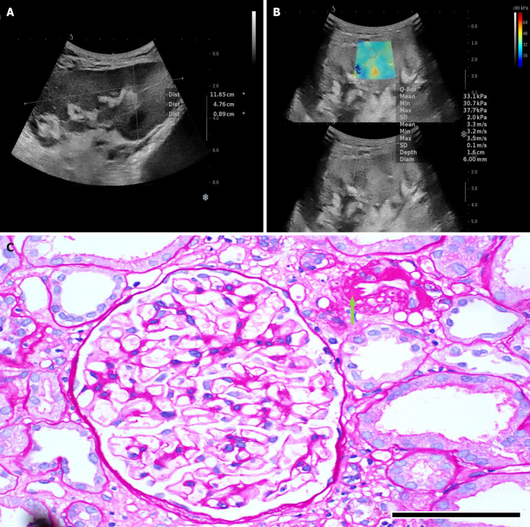
The study focuses on the use of multi-parametric ultrasound [gray scale, color Doppler and shear wave elastography (SWE)] to differentiate stable renal allografts from acute graft dysfunction and to assess time-dependent changes in parenchymal stiffness, thereby assessing its use as an efficient monitoring tool for ongoing graft dysfunction. To date, biopsy is the gold standard for evaluation of acute graft dysfunction. However, because it is invasive, it carries certain risks and cannot be used for follow-up monitoring. SWE is a non-invasive imaging modality that identifies higher parenchymal stiffness values in cases of acute graft dysfunction compared to stable grafts.

AIM

To assess renal allograft parenchymal stiffness by SWE and to correlate its findings with functional status of the graft kidney.

METHODS

This prospective observational study included 71 renal allograft recipients. Multi-parametric ultrasound was performed on all patients, and biopsies were performed in cases of acute graft dysfunction. The study was performed for a period of 2 years at Sanjay Gandhi Postgraduate Institute of Medical Sciences, Lucknow, a tertiary care center in north India. Independent samples -test was used to compare the means between two independent groups. Paired-samples -test was used to test the change in mean value between baseline and follow-up observations.

RESULTS

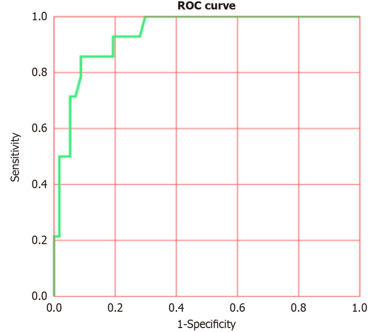
Thirty-one patients had experienced acute graft dysfunction at least once, followed by recovery, but none of them had a history of chronic renal allograft injury. Mean baseline parenchymal stiffness in stable grafts and acute graft dysfunction were 30.21 + 2.03 kPa (3.17 + 0.11 m/s) and 31.07 + 2.88 kPa (3.22 + 0.15 m/s), respectively; however, these differences were not statistically significant ( = 0.305 and 0.252, respectively). There was a gradual decrease in SWE values during the first 3 postoperative months, followed by an increase in SWE values up to one-year post-transplantation. Patients with biopsy-confirmed graft dysfunction showed higher SWE values compared to those with a negative biopsy. However, receiver operating characteristic analysis failed to show statistically significant cut-off values to differentiate between the stable graft and acute graft dysfunction.

CONCLUSION

Acute graft dysfunction displays higher parenchymal stiffness values compared to stable grafts. Therefore, SWE may be useful in monitoring the functional status of allografts to predict any ongoing dysfunction.

摘要

背景

本研究聚焦于运用多参数超声[灰阶、彩色多普勒及剪切波弹性成像(SWE)]来区分稳定的肾移植与急性移植肾功能障碍,并评估实质硬度随时间的变化,从而评估其作为持续移植肾功能障碍有效监测工具的用途。迄今为止,活检是评估急性移植肾功能障碍的金标准。然而,由于其具有侵入性,存在一定风险且不能用于随访监测。SWE是一种非侵入性成像方式,与稳定移植相比,其在急性移植肾功能障碍病例中可识别出更高的实质硬度值。

目的

通过SWE评估肾移植实质硬度,并将其结果与移植肾的功能状态相关联。

方法

这项前瞻性观察性研究纳入了71名肾移植受者。对所有患者进行了多参数超声检查,对于急性移植肾功能障碍患者进行了活检。该研究在印度北部三级医疗中心勒克瑙的桑贾伊·甘地医学科学研究生学院进行了2年。采用独立样本t检验比较两个独立组之间的均值。采用配对样本t检验来检验基线与随访观察之间均值的变化。

结果

31名患者至少经历过一次急性移植肾功能障碍,随后恢复,但他们均无慢性肾移植损伤史。稳定移植组和急性移植肾功能障碍组的平均基线实质硬度分别为30.21 + 2.03 kPa(3.17 + 0.11 m/s)和31.07 + 2.88 kPa(3.22 + 0.15 m/s);然而,这些差异无统计学意义(分别为 = 0.305和0.252)。术后前3个月SWE值逐渐下降,随后直至移植后1年SWE值升高。活检证实有移植肾功能障碍的患者与活检阴性的患者相比,SWE值更高。然而,受试者工作特征分析未能显示出区分稳定移植与急性移植肾功能障碍的具有统计学意义的临界值。

结论

与稳定移植相比,急性移植肾功能障碍表现出更高的实质硬度值。因此,SWE可能有助于监测移植肾的功能状态以预测任何持续的功能障碍。

https://cdn.ncbi.nlm.nih.gov/pmc/blobs/1938/11718520/eb35ba83308f/WJR-16-782-g007.jpg
https://cdn.ncbi.nlm.nih.gov/pmc/blobs/1938/11718520/8d4b291296bb/WJR-16-782-g001.jpg
https://cdn.ncbi.nlm.nih.gov/pmc/blobs/1938/11718520/7d9213ec3daf/WJR-16-782-g002.jpg
https://cdn.ncbi.nlm.nih.gov/pmc/blobs/1938/11718520/2c1334c5c2e8/WJR-16-782-g003.jpg
https://cdn.ncbi.nlm.nih.gov/pmc/blobs/1938/11718520/7922c5e648a0/WJR-16-782-g004.jpg
https://cdn.ncbi.nlm.nih.gov/pmc/blobs/1938/11718520/607eae7e67ec/WJR-16-782-g005.jpg
https://cdn.ncbi.nlm.nih.gov/pmc/blobs/1938/11718520/3b1269f4dbd2/WJR-16-782-g006.jpg
https://cdn.ncbi.nlm.nih.gov/pmc/blobs/1938/11718520/eb35ba83308f/WJR-16-782-g007.jpg

相似文献

1
Role of multi-parametric ultrasonography for the assessment and monitoring of functional status of renal allografts with histopathological correlation.多参数超声检查在评估和监测肾移植功能状态及与组织病理学相关性方面的作用
World J Radiol. 2024 Dec 28;16(12):782-793. doi: 10.4329/wjr.v16.i12.782.
2
Role of Shear Wave Elastography for Assessment of Renal-Allograft Fibrosis and its Correlation With Histopathology.剪切波弹性成像在评估肾移植纤维化及其与组织病理学相关性中的作用。
J Ultrasound Med. 2024 Oct;43(10):1979-1992. doi: 10.1002/jum.16532. Epub 2024 Jul 30.
3
ARFI-based tissue elasticity quantification and kidney graft dysfunction: first clinical experiences.基于声辐射力脉冲成像的组织弹性定量分析与移植肾失功:初步临床经验
Clin Hemorheol Microcirc. 2011;49(1-4):527-35. doi: 10.3233/CH-2011-1503.
4
Renal Allograft Dysfunction: Evaluation with Shear-wave Sonoelastography.肾移植功能障碍:剪切波弹性成像评估。
Radiology. 2018 Jul;288(1):146-152. doi: 10.1148/radiol.2018170577. Epub 2018 Apr 10.
5
Prediction of acute rejection in renal allografts using shear-wave dispersion slope.利用剪切波频散斜率预测肾移植急性排斥反应
Eur Radiol. 2024 Jul;34(7):4527-4537. doi: 10.1007/s00330-023-10492-8. Epub 2023 Dec 13.
6
Shear-Wave Elastography Variability Analysis and Relation with Kidney Allograft Dysfunction: A Single-Center Study.剪切波弹性成像变异性分析及其与肾移植功能障碍的关系:一项单中心研究
Diagnostics (Basel). 2020 Jan 13;10(1):41. doi: 10.3390/diagnostics10010041.
7
Ultrasound Shear Wave Elastography for Liver Disease. A Critical Appraisal of the Many Actors on the Stage.用于肝脏疾病的超声剪切波弹性成像:对该领域众多参与者的批判性评估
Ultraschall Med. 2016 Feb;37(1):1-5. doi: 10.1055/s-0035-1567037. Epub 2016 Feb 12.
8
Application value of shear-wave elastography combined with monochrome superb microvascular imaging in renal allograft chronic rejection.剪切波弹性成像联合单色超微血管成像在肾移植慢性排斥反应中的应用价值
Clin Hemorheol Microcirc. 2022;82(4):303-311. doi: 10.3233/CH-221443.
9
Non-invasive evaluation of stable renal allograft function using point shear-wave elastography.使用点剪切波弹性成像技术对稳定肾移植功能进行无创评估。
Br J Radiol. 2018 Jan;91(1081):20170372. doi: 10.1259/bjr.20170372. Epub 2017 Oct 31.
10
Utility of Shear Wave Elastography for Assessing Allograft Fibrosis in Renal Transplant Recipients: A Pilot Study.剪切波弹性成像在评估肾移植受者同种异体移植纤维化中的应用:一项初步研究。
J Ultrasound Med. 2018 Jun;37(6):1455-1465. doi: 10.1002/jum.14487. Epub 2017 Nov 16.

引用本文的文献

1
Update on newer ultrasound systems to study the microvasculature.用于研究微血管系统的新型超声系统的最新进展。
Radiol Med. 2025 Jun 25. doi: 10.1007/s11547-025-02035-6.

本文引用的文献

1
Shear wave elastography and Doppler ultrasound in kidney transplant recipients.肾移植受者的剪切波弹性成像与多普勒超声
Radiol Bras. 2022 Jan-Feb;55(1):19-23. doi: 10.1590/0100-3984.2020.0148.
2
Evaluation of the Graft Kidney in the Early Postoperative Period: Performance of Contrast-Enhanced Ultrasound and Additional Ultrasound Parameters.移植肾术后早期评估:对比增强超声及附加超声参数的应用。
J Ultrasound Med. 2021 Sep;40(9):1771-1783. doi: 10.1002/jum.15557. Epub 2020 Nov 12.
3
Shear-Wave Elastography Variability Analysis and Relation with Kidney Allograft Dysfunction: A Single-Center Study.
剪切波弹性成像变异性分析及其与肾移植功能障碍的关系:一项单中心研究
Diagnostics (Basel). 2020 Jan 13;10(1):41. doi: 10.3390/diagnostics10010041.
4
Update on imaging-based diagnosis of acute renal allograft rejection.基于影像学的急性肾移植排斥反应诊断的最新进展。
Am J Nucl Med Mol Imaging. 2019 Apr 15;9(2):110-126. eCollection 2019.
5
Renal Allograft Dysfunction: Evaluation with Shear-wave Sonoelastography.肾移植功能障碍:剪切波弹性成像评估。
Radiology. 2018 Jul;288(1):146-152. doi: 10.1148/radiol.2018170577. Epub 2018 Apr 10.
6
Utility of Shear Wave Elastography for Assessing Allograft Fibrosis in Renal Transplant Recipients: A Pilot Study.剪切波弹性成像在评估肾移植受者同种异体移植纤维化中的应用:一项初步研究。
J Ultrasound Med. 2018 Jun;37(6):1455-1465. doi: 10.1002/jum.14487. Epub 2017 Nov 16.
7
Non-invasive evaluation of stable renal allograft function using point shear-wave elastography.使用点剪切波弹性成像技术对稳定肾移植功能进行无创评估。
Br J Radiol. 2018 Jan;91(1081):20170372. doi: 10.1259/bjr.20170372. Epub 2017 Oct 31.
8
Usefulness of Multiparametric Ultrasound for Evaluating Structural Abnormality of Transplanted Kidney: Can We Predict Histologic Abnormality on Renal Biopsy in Advance?多参数超声评估移植肾结构异常的实用性:我们能否提前预测肾活检的组织学异常?
AJR Am J Roentgenol. 2017 Sep;209(3):W139-W144. doi: 10.2214/AJR.16.17397. Epub 2017 Jul 13.
9
Strategies for long-term preservation of kidney graft function.肾脏移植物功能的长期保护策略。
Lancet. 2017 May 27;389(10084):2152-2162. doi: 10.1016/S0140-6736(17)31283-7.
10
Challenges and Considerations When Using Shear Wave Elastography to Evaluate the Transplanted Kidney, With Pictorial Review.使用剪切波弹性成像评估移植肾的挑战与注意事项,并附图片回顾
J Ultrasound Med. 2017 Sep;36(9):1771-1782. doi: 10.1002/jum.14217. Epub 2017 May 4.