Cabeza-Gil Iulen, Ruggeri Marco, Manns Fabrice
Aragón Institute of Engineering Research (i3A), University of Zaragoza, Zaragoza, Spain.
Ophthalmic Biophysics Center, Bascom Palmer Eye Institute, University of Miami Miller School of Medicine, Miami, FL, USA.
Transl Vis Sci Technol. 2025 Jan 2;14(1):17. doi: 10.1167/tvst.14.1.17.
Although the lens undoubtedly plays a major role in presbyopia, altered lens function could be in part secondary to age-related changes of the ciliary muscle. Ciliary muscle changes with accommodation have been quantified using optical coherence tomography, but so far these studies have been limited to quantifying changes in ciliary muscle thickness, mostly at static accommodative states. Quantifying ciliary muscle thickness changes does not effectively capture the dynamic anterior-centripetal movement of the ciliary muscle during accommodation. To address this issue, we present a method to quantify the movement of the ciliary muscle during accommodation using trans-scleral optical coherence tomography images obtained dynamically.
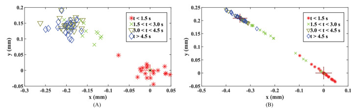
An image processing framework including distortion correction, geometric transformation, and Procrustes analysis, was used to quantify the anterior-centripetal movement of the ciliary muscle apex and centroid during accommodation. The method was applied in a preliminary study to quantify ciliary muscle displacement and its relation to lens thickness change with accommodation on two young adults and two prepresbyopes.
The magnitude and the direction relative to the pupil plane of the apex/centroid displacement in response to a two diopters (2D) stimulus were 0.16/0.20 mm at 11.3°/30.5° and 0.26/0.34 mm at 6.6°/33.2° for the young adults and 0.20/0.20 mm at 29.7°/40.6° and 0.24/0.40 mm at 33.0°/31.7° for the prepresbyopes, respectively.
This study demonstrates the feasibility of quantifying dynamic anterior-centripetal movement of the ciliary muscle during accommodation using optical coherence tomography. The method better captures the functional response of the muscle than the quantification of thickness changes.
We provide a method that holds potential to better understand the age-related changes of the ciliary muscle on presbyopia.
尽管晶状体在老花眼中无疑起着主要作用,但晶状体功能改变可能部分继发于睫状肌的年龄相关性变化。已使用光学相干断层扫描对睫状肌随调节的变化进行了量化,但到目前为止,这些研究仅限于量化睫状肌厚度的变化,主要是在静态调节状态下。量化睫状肌厚度变化并不能有效地捕捉调节过程中睫状肌的动态向心运动。为了解决这个问题,我们提出了一种方法,使用动态获得的经巩膜光学相干断层扫描图像来量化调节过程中睫状肌的运动。
使用包括畸变校正、几何变换和普氏分析的图像处理框架,来量化调节过程中睫状肌顶点和质心的向心运动。该方法应用于一项初步研究,以量化两名年轻成年人和两名早老性远视患者调节时睫状肌的位移及其与晶状体厚度变化的关系。
对于年轻成年人,在2屈光度(2D)刺激下,顶点/质心位移相对于瞳孔平面的大小和方向分别为11.3°/30.5°时为0.16/0.20毫米,6.6°/33.2°时为0.26/0.34毫米;对于早老性远视患者,分别为29.7°/40.6°时为0.20/0.20毫米,33.0°/31.7°时为0.24/0.40毫米。
本研究证明了使用光学相干断层扫描量化调节过程中睫状肌动态向心运动的可行性。该方法比量化厚度变化能更好地捕捉肌肉的功能反应。
我们提供了一种方法,有可能更好地理解睫状肌与老花眼相关的年龄相关性变化。