Özen Ilknur, Hamdeh Sami Abu, Ruscher Karsten, Marklund Niklas
Department of Clinical Sciences, Lund Brain Injury Laboratory for Neurosurgical Research, Lund University, 222 20, Lund, Sweden.
Department of Medical Sciences, Section of Neurosurgery, Uppsala University, Uppsala, Sweden.
Acta Neuropathol. 2025 Jan 22;149(1):10. doi: 10.1007/s00401-025-02848-9.
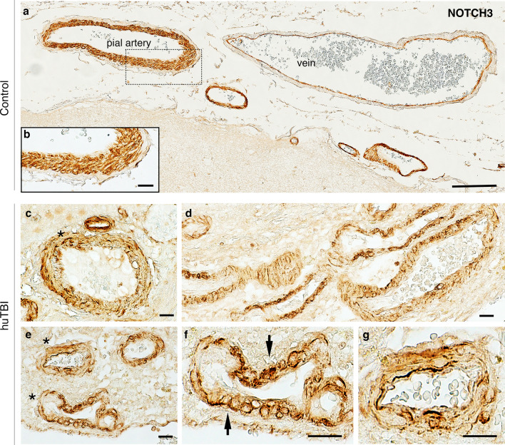
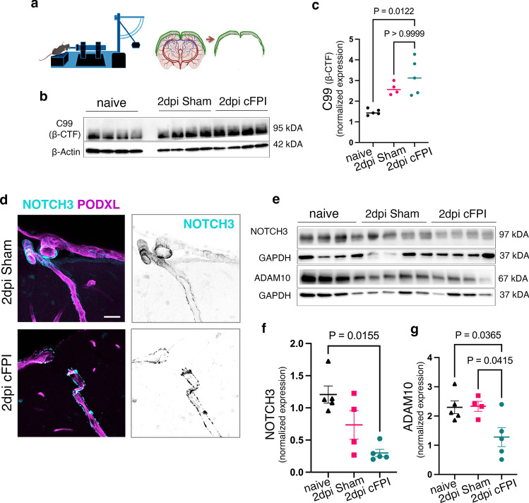
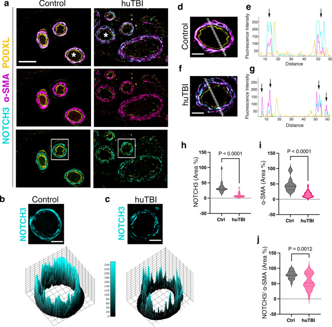
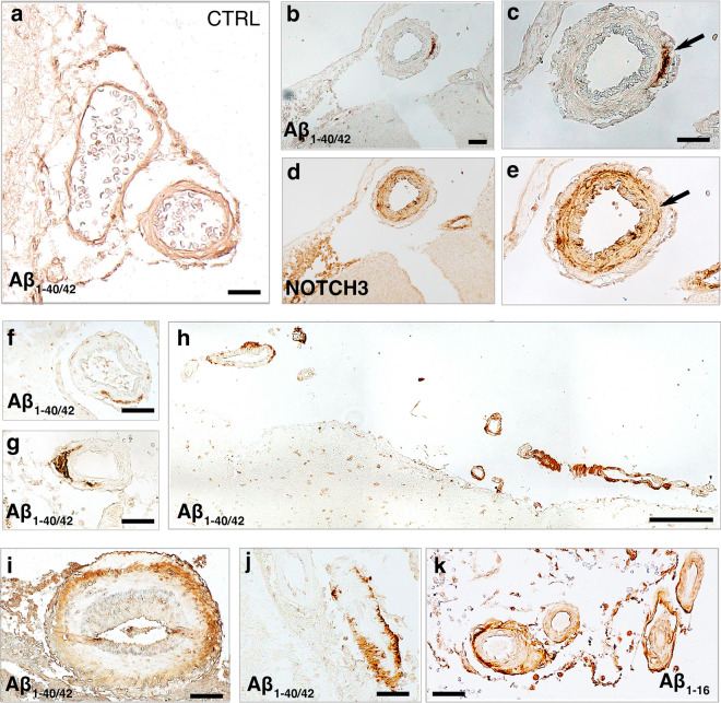
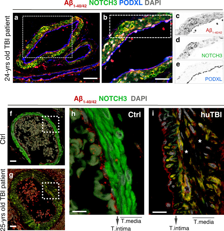
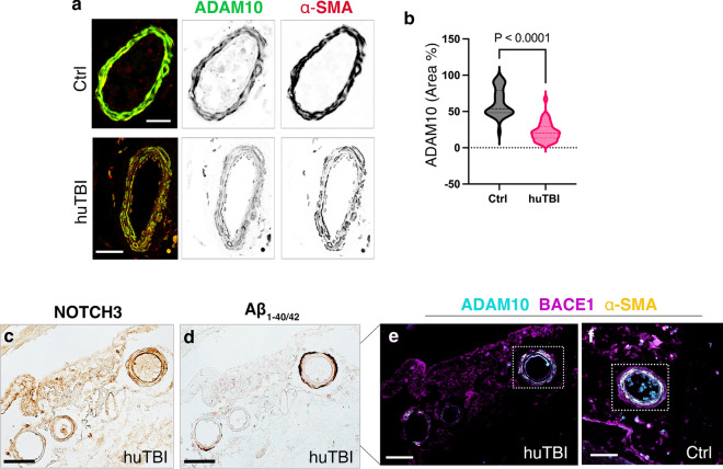
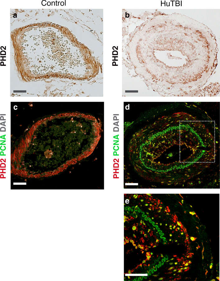
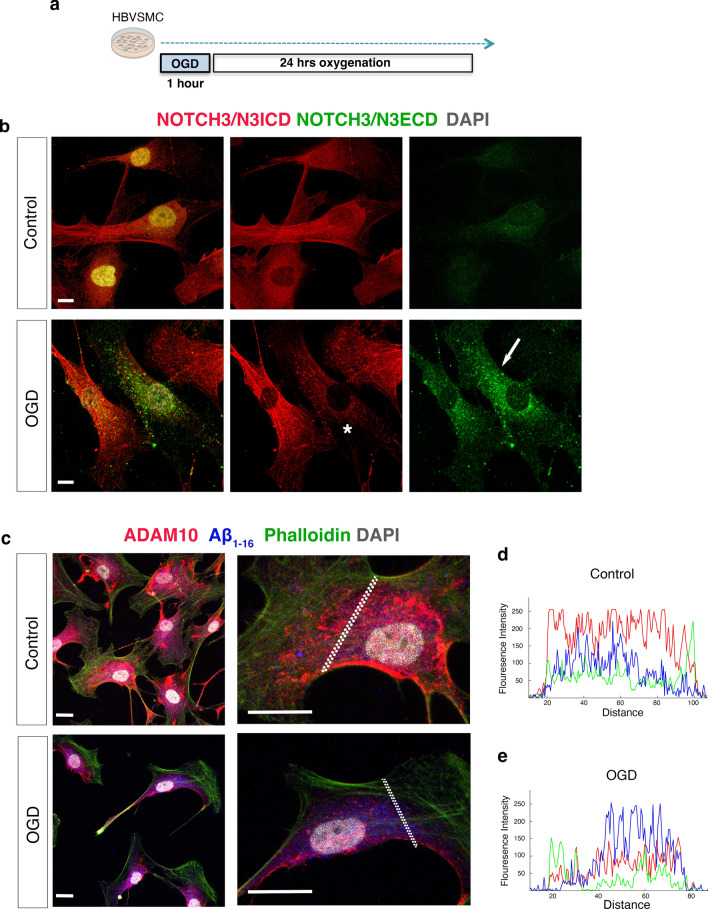
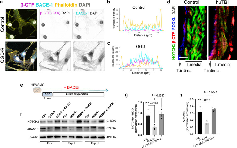
Traumatic brain injury (TBI) often leads to impaired regulation of cerebral blood flow, which may be caused by pathological changes of the vascular smooth muscle cells (VSMCs) in the arterial wall. Moreover, these cerebrovascular changes may contribute to the development of various neurodegenerative disorders such as Alzheimer's-like pathologies that include amyloid beta aggregation. Despite its importance, the pathophysiological mechanisms responsible for VSMC dysfunction after TBI have rarely been evaluated. Here, we show that acute human TBI resulted in early pathological changes in leptomeningeal arteries, closely associated with a decrease in VSMC markers such as NOTCH3 and alpha smooth muscle actin (α-SMA).These changes coincided with increased aggregation of variable-length amyloid peptides including Aβ Aβ and β-secretase-derived fragment (βCTF) (C99) caused by altered processing of amyloid precursor protein (APP) in VSMCs. The aggregation of Aβ peptides were also observed in the leptomeningeal arteries of young TBI patients. These pathological changes also included higher β-secretase (BACE1) when compared to α-secretase A Disintegrin And Metalloprotease 10 (ADAM10) expression in the leptomeningeal arteries, plausibly caused by hypoxia and oxidative stress as shown using human VSMCs in vitro. Importantly, BACE1 inhibition not only restored NOTCH3 signalling but also normalized ADAM10 levels in vitro. Furthermore, we found reduced ADAM10 activity and decreased NOTCH3, along with increased βCTF (C99) levels in mice subjected to an experimental model of TBI. This study provides evidence of early post-injury changes in VSMCs of leptomeningeal arteries that can contribute to vascular dysfunction and exacerbate secondary injury mechanisms following TBI.
创伤性脑损伤(TBI)常导致脑血流调节受损,这可能由动脉壁血管平滑肌细胞(VSMC)的病理变化引起。此外,这些脑血管变化可能促使各种神经退行性疾病的发展,如包括淀粉样β蛋白聚集的阿尔茨海默氏样病变。尽管其重要性,但TBI后导致VSMC功能障碍的病理生理机制很少得到评估。在此,我们表明急性人类TBI导致软脑膜动脉早期病理变化,这与VSMC标志物如NOTCH3和α平滑肌肌动蛋白(α-SMA)的减少密切相关。这些变化与可变长度淀粉样肽包括Aβ和β-分泌酶衍生片段(βCTF)(C99)的聚集增加同时发生,这是由VSMC中淀粉样前体蛋白(APP)加工改变所致。在年轻TBI患者的软脑膜动脉中也观察到Aβ肽的聚集。与软脑膜动脉中α-分泌酶解整合素和金属蛋白酶10(ADAM10)的表达相比,这些病理变化还包括更高的β-分泌酶(BACE1)表达,体外使用人类VSMC显示这可能由缺氧和氧化应激引起。重要的是,BACE1抑制不仅恢复了体外NOTCH3信号传导,还使ADAM10水平正常化。此外,我们发现实验性TBI小鼠中ADAM10活性降低、NOTCH3减少以及βCTF(C99)水平升高。本研究提供了软脑膜动脉VSMC损伤后早期变化的证据,这些变化可导致血管功能障碍并加剧TBI后的继发性损伤机制。