• 文献检索
  • 文档翻译
  • 深度研究
  • 学术资讯
  • Suppr Zotero 插件Zotero 插件
  • 邀请有礼
  • 套餐&价格
  • 历史记录
应用&插件
Suppr Zotero 插件Zotero 插件浏览器插件Mac 客户端Windows 客户端微信小程序
定价
高级版会员购买积分包购买API积分包
服务
文献检索文档翻译深度研究API 文档MCP 服务
关于我们
关于 Suppr公司介绍联系我们用户协议隐私条款
关注我们

Suppr 超能文献

核心技术专利:CN118964589B侵权必究
粤ICP备2023148730 号-1Suppr @ 2026

文献检索

告别复杂PubMed语法,用中文像聊天一样搜索,搜遍4000万医学文献。AI智能推荐,让科研检索更轻松。

立即免费搜索

文件翻译

保留排版,准确专业,支持PDF/Word/PPT等文件格式,支持 12+语言互译。

免费翻译文档

深度研究

AI帮你快速写综述,25分钟生成高质量综述,智能提取关键信息,辅助科研写作。

立即免费体验

一个带注释的异构超声数据库。

An annotated heterogeneous ultrasound database.

作者信息

Yang Yuezhe, Chen Yonglin, Dong Xingbo, Zhang Junning, Long Chihui, Jin Zhe, Dai Yong

机构信息

Anhui Provincial International Joint Research Center for Advanced Technology in Medical Imaging, School of Artificial Intelligence, Anhui University, Hefei, 230601, China.

School of Electronic and Information Engineering, Anhui Jianzhu University, Hefei, 230601, China.

出版信息

Sci Data. 2025 Jan 25;12(1):148. doi: 10.1038/s41597-025-04464-4.

DOI:10.1038/s41597-025-04464-4
PMID:39863639
原文链接:https://pmc.ncbi.nlm.nih.gov/articles/PMC11762285/
Abstract

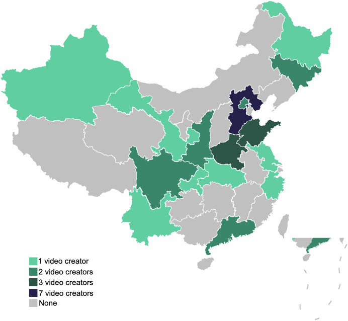
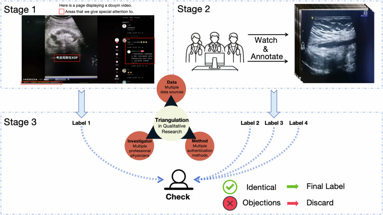
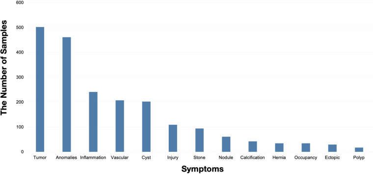
Ultrasound is a primary diagnostic tool commonly used to evaluate internal body structures, including organs, blood vessels, the musculoskeletal system, and fetal development. Due to challenges such as operator dependence, noise, limited field of view, difficulty in imaging through bone and air, and variability across different systems, diagnosing abnormalities in ultrasound images is particularly challenging for less experienced clinicians. The development of artificial intelligence (AI) technology could assist in the diagnosis of ultrasound images. However, many databases are created using a single device type and collection site, limiting the generalizability of machine learning models. Therefore, we have collected a large, publicly accessible ultrasound challenge database that is intended to significantly enhance the performance of AI-assisted ultrasound diagnosis. This database is derived from publicly available data on the Internet and comprises a total of 1,833 distinct ultrasound data. It includes 13 different ultrasound image anomalies, and all data have been anonymized. Our data-sharing program aims to support benchmark testing of ultrasound disease diagnosis in multi-center environments.

摘要

超声是一种常用的主要诊断工具,用于评估体内结构,包括器官、血管、肌肉骨骼系统和胎儿发育情况。由于存在诸如依赖操作人员、噪声、视野有限、难以透过骨骼和空气成像以及不同系统之间存在差异等挑战,对于经验不足的临床医生而言,在超声图像中诊断异常情况极具挑战性。人工智能(AI)技术的发展有助于超声图像的诊断。然而,许多数据库是使用单一设备类型和采集地点创建的,这限制了机器学习模型的通用性。因此,我们收集了一个大型的、可公开访问的超声挑战数据库,旨在显著提高人工智能辅助超声诊断的性能。该数据库源自互联网上的公开可用数据,总共包含1833个不同的超声数据。它包括13种不同的超声图像异常情况,并且所有数据均已匿名化。我们的数据共享计划旨在支持多中心环境下超声疾病诊断的基准测试。

https://cdn.ncbi.nlm.nih.gov/pmc/blobs/8da5/11762285/9681592b137e/41597_2025_4464_Fig5_HTML.jpg
https://cdn.ncbi.nlm.nih.gov/pmc/blobs/8da5/11762285/4aa54a60fcc1/41597_2025_4464_Fig1_HTML.jpg
https://cdn.ncbi.nlm.nih.gov/pmc/blobs/8da5/11762285/ae1a1f502f19/41597_2025_4464_Fig2_HTML.jpg
https://cdn.ncbi.nlm.nih.gov/pmc/blobs/8da5/11762285/34ba28dbc47e/41597_2025_4464_Fig3_HTML.jpg
https://cdn.ncbi.nlm.nih.gov/pmc/blobs/8da5/11762285/1b1982bb62d7/41597_2025_4464_Fig4_HTML.jpg
https://cdn.ncbi.nlm.nih.gov/pmc/blobs/8da5/11762285/9681592b137e/41597_2025_4464_Fig5_HTML.jpg

相似文献

1
An annotated heterogeneous ultrasound database.一个带注释的异构超声数据库。
Sci Data. 2025 Jan 25;12(1):148. doi: 10.1038/s41597-025-04464-4.
2
Artificial intelligence in ultrasound.人工智能在超声中的应用。
Eur J Radiol. 2021 Jun;139:109717. doi: 10.1016/j.ejrad.2021.109717. Epub 2021 Apr 12.
3
Intelligent diagnosis of thyroid nodules with AI ultrasound assistance and cytology classification.借助人工智能超声辅助和细胞学分类对甲状腺结节进行智能诊断。
Front Endocrinol (Lausanne). 2025 May 21;16:1546983. doi: 10.3389/fendo.2025.1546983. eCollection 2025.
4
Interaction between clinicians and artificial intelligence to detect fetal atrioventricular septal defects on ultrasound: how can we optimize collaborative performance?超声检测胎儿房室间隔缺损中临床医生与人工智能的相互作用:如何优化协作表现?
Ultrasound Obstet Gynecol. 2024 Jul;64(1):28-35. doi: 10.1002/uog.27577. Epub 2024 Jun 3.
5
Artificial Intelligence: Innovation to Assist in the Identification of Sono-anatomy for Ultrasound-Guided Regional Anaesthesia.人工智能:创新以协助超声引导区域麻醉中的 Sono 解剖学识别。
Adv Exp Med Biol. 2022;1356:117-140. doi: 10.1007/978-3-030-87779-8_6.
6
Prenatal detection of congenital heart defects using the deep learning-based image and video analysis: protocol for Clinical Artificial Intelligence in Fetal Echocardiography (CAIFE), an international multicentre multidisciplinary study.使用基于深度学习的图像和视频分析进行先天性心脏缺陷的产前检测:胎儿超声心动图临床人工智能(CAIFE)方案,一项国际多中心多学科研究。
BMJ Open. 2025 Jun 5;15(6):e101263. doi: 10.1136/bmjopen-2025-101263.
7
Artificial Intelligence-enabled, Real-time Intraoperative Ultrasound Imaging of Neural Structures Within the Psoas: Validation in a Porcine Spine Model.人工智能辅助实时超声成像在腰大肌内神经结构中的应用:在猪脊柱模型中的验证。
Spine (Phila Pa 1976). 2021 Feb 1;46(3):E146-E152. doi: 10.1097/BRS.0000000000003704.
8
Transfer learning for accurate fetal organ classification from ultrasound images: a potential tool for maternal healthcare providers.从超声图像中进行准确的胎儿器官分类的迁移学习:孕产妇医疗保健提供者的潜在工具。
Sci Rep. 2023 Oct 20;13(1):17904. doi: 10.1038/s41598-023-44689-0.
9
Use of Artificial Intelligence and Machine Learning in Critical Care Ultrasound.人工智能和机器学习在重症监护超声中的应用。
Crit Care Clin. 2025 Jul;41(3):593-608. doi: 10.1016/j.ccc.2025.02.008. Epub 2025 Apr 7.
10
Diagnostic Performance of an Artificial Intelligence System in Breast Ultrasound.人工智能系统在乳腺超声中的诊断性能。
J Ultrasound Med. 2022 Jan;41(1):97-105. doi: 10.1002/jum.15684. Epub 2021 Mar 5.

引用本文的文献

1
TN5000: An Ultrasound Image Dataset for Thyroid Nodule Detection and Classification.TN5000:用于甲状腺结节检测与分类的超声图像数据集。
Sci Data. 2025 Aug 16;12(1):1437. doi: 10.1038/s41597-025-05757-4.

本文引用的文献

1
An ultrasonography of thyroid nodules dataset with pathological diagnosis annotation for deep learning.甲状腺结节超声数据集,附有深度学习用的病理诊断标注。
Sci Data. 2024 Nov 23;11(1):1272. doi: 10.1038/s41597-024-04156-5.
2
Attention based UNet model for breast cancer segmentation using BUSI dataset.基于注意力机制的 UNet 模型在 BUSI 数据集上的乳腺癌分割
Sci Rep. 2024 Sep 28;14(1):22422. doi: 10.1038/s41598-024-72712-5.
3
PSFHS: Intrapartum ultrasound image dataset for AI-based segmentation of pubic symphysis and fetal head.
PSFHS:基于人工智能的耻骨联合和胎儿头部分割的产时超声图像数据集。
Sci Data. 2024 May 2;11(1):436. doi: 10.1038/s41597-024-03266-4.
4
Multimodal Deep Learning for Integrating Chest Radiographs and Clinical Parameters: A Case for Transformers.多模态深度学习在整合胸部 X 光片和临床参数中的应用:基于变压器的方法。
Radiology. 2023 Oct;309(1):e230806. doi: 10.1148/radiol.230806.
5
Deep Long-Tailed Learning: A Survey.深度长尾学习:一项综述。
IEEE Trans Pattern Anal Mach Intell. 2023 Sep;45(9):10795-10816. doi: 10.1109/TPAMI.2023.3268118. Epub 2023 Aug 7.
6
A review on deep-learning algorithms for fetal ultrasound-image analysis.胎儿超声图像分析的深度学习算法综述
Med Image Anal. 2023 Jan;83:102629. doi: 10.1016/j.media.2022.102629. Epub 2022 Oct 14.
7
Extracting keyframes of breast ultrasound video using deep reinforcement learning.使用深度强化学习提取乳腺超声视频的关键帧。
Med Image Anal. 2022 Aug;80:102490. doi: 10.1016/j.media.2022.102490. Epub 2022 Jun 5.
8
Current challenges of implementing artificial intelligence in medical imaging.当前在医学影像中实施人工智能所面临的挑战。
Phys Med. 2022 Aug;100:12-17. doi: 10.1016/j.ejmp.2022.06.003. Epub 2022 Jun 14.
9
Learning From Noisy Labels With Deep Neural Networks: A Survey.基于深度神经网络从噪声标签中学习:一项综述。
IEEE Trans Neural Netw Learn Syst. 2023 Nov;34(11):8135-8153. doi: 10.1109/TNNLS.2022.3152527. Epub 2023 Oct 27.
10
Hierarchical deep learning models using transfer learning for disease detection and classification based on small number of medical images.基于少量医学图像的疾病检测和分类的迁移学习的分层深度学习模型。
Sci Rep. 2021 Mar 1;11(1):4250. doi: 10.1038/s41598-021-83503-7.