Akbari Faezeh, Liu Xuhui, Hamedi Fatemeh, Mohtasebi Mehrana, Chen Li, Chen Lei, Yu Guoqiang
University of Kentucky, Department of Biomedical Engineering, Lexington, Kentucky, United States.
University of Kentucky, Biostatistics and Bioinformatics Shared Resource Facility, Markey Cancer Center, Lexington, Kentucky, United States.
Neurophotonics. 2025 Jan;12(1):015006. doi: 10.1117/1.NPh.12.1.015006. Epub 2025 Jan 27.
Cerebral blood flow (CBF) imaging is crucial for diagnosing cerebrovascular diseases. However, existing large neuroimaging techniques with high cost, low sampling rate, and poor mobility make them unsuitable for continuous and longitudinal CBF monitoring at the bedside.
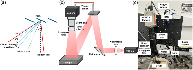
We aimed to develop a low-cost, portable, programmable scanning diffuse speckle contrast imaging (PS-DSCI) technology for fast, high-density, and depth-sensitive imaging of CBF in rodents.
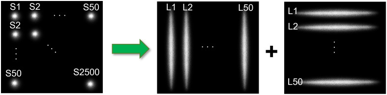
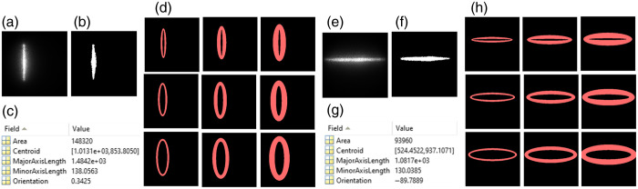
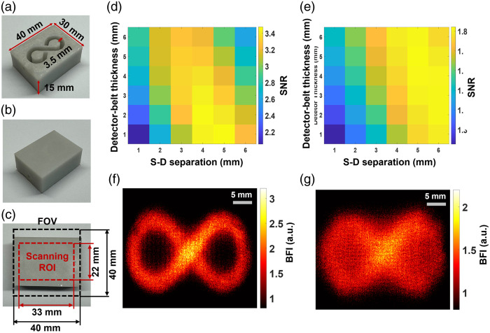
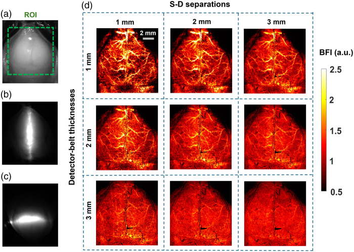
The PS-DSCI employed a programmable digital micromirror device (DMD) for remote line-shaped laser (785 nm) scanning on tissue surface and synchronized a 2D camera for capturing boundary diffuse laser speckle contrasts. New algorithms were developed to address deformations of line-shaped scanning, thus minimizing CBF reconstruction artifacts. The PS-DSCI was examined in head-simulating phantoms and adult mice.
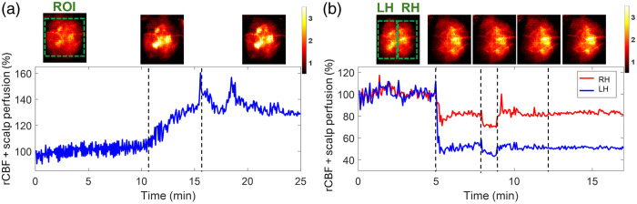
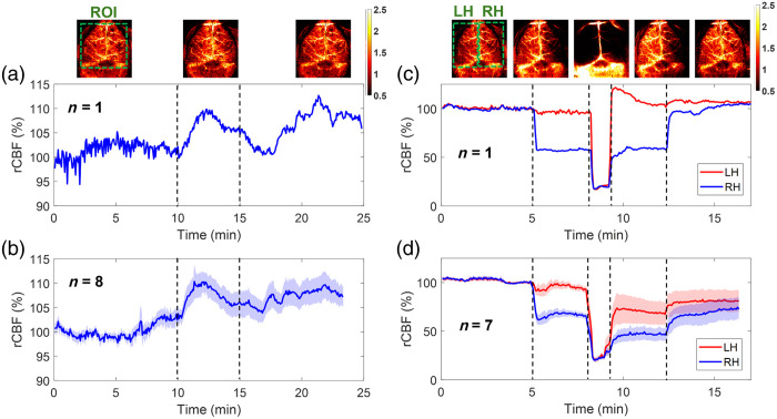
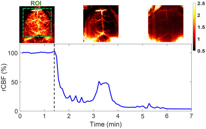
The PS-DSCI enables resolving intralipid particle flow contrasts at different tissue depths. experiments in adult mice demonstrated the capability of PS-DSCI to image global/regional CBF variations induced by 8% inhalation and transient carotid artery ligations.
Compared with conventional point scanning, line scanning in PS-DSCI significantly increases spatiotemporal resolution. The high sampling rate of PS-DSCI is crucial for capturing rapid CBF changes while high spatial resolution is important for visualizing brain vasculature.
脑血流(CBF)成像对于诊断脑血管疾病至关重要。然而,现有的大型神经成像技术成本高、采样率低且移动性差,使其不适合在床边进行连续和纵向的CBF监测。
我们旨在开发一种低成本、便携式、可编程扫描漫散斑对比成像(PS-DSCI)技术,用于对啮齿动物的CBF进行快速、高密度和深度敏感成像。
PS-DSCI采用可编程数字微镜器件(DMD)在组织表面进行远程线状激光(785nm)扫描,并同步使用二维相机捕获边界漫散激光散斑对比度。开发了新算法来解决线状扫描的变形问题,从而最大限度地减少CBF重建伪影。在头部模拟体模和成年小鼠中对PS-DSCI进行了检测。
PS-DSCI能够分辨不同组织深度的脂质内颗粒流动对比度。在成年小鼠中的实验证明了PS-DSCI对由8%吸入和短暂颈动脉结扎引起的全局/区域CBF变化进行成像的能力。
与传统点扫描相比,PS-DSCI中的线扫描显著提高了时空分辨率。PS-DSCI的高采样率对于捕获快速的CBF变化至关重要,而高空间分辨率对于可视化脑血管系统很重要。