• 文献检索
  • 文档翻译
  • 深度研究
  • 学术资讯
  • Suppr Zotero 插件Zotero 插件
  • 邀请有礼
  • 套餐&价格
  • 历史记录
应用&插件
Suppr Zotero 插件Zotero 插件浏览器插件Mac 客户端Windows 客户端微信小程序
定价
高级版会员购买积分包购买API积分包
服务
文献检索文档翻译深度研究API 文档MCP 服务
关于我们
关于 Suppr公司介绍联系我们用户协议隐私条款
关注我们

Suppr 超能文献

核心技术专利:CN118964589B侵权必究
粤ICP备2023148730 号-1Suppr @ 2026

文献检索

告别复杂PubMed语法,用中文像聊天一样搜索,搜遍4000万医学文献。AI智能推荐,让科研检索更轻松。

立即免费搜索

文件翻译

保留排版,准确专业,支持PDF/Word/PPT等文件格式,支持 12+语言互译。

免费翻译文档

深度研究

AI帮你快速写综述,25分钟生成高质量综述,智能提取关键信息,辅助科研写作。

立即免费体验

四种基于深度学习的腹部危险器官自动分割商用软件的评估与故障分析

Evaluation and failure analysis of four commercial deep learning-based autosegmentation software for abdominal organs at risk.

作者信息

Fan Mingdong, Wang Tonghe, Lei Yang, Patel Pretesh R, Dresser Sean, Ghavidel Beth Bradshaw, Qiu Richard L J, Zhou Jun, Luca Kirk, Kayode Oluwatosin, Bradley Jeffrey D, Yang Xiaofeng, Roper Justin

机构信息

Department of Radiation Oncology and Winship Cancer Institute, Emory University, Atlanta, Georgia, USA.

Department of Medical Physics, Memorial Sloan Kettering Cancer Center, New York, New York, USA.

出版信息

J Appl Clin Med Phys. 2025 Apr;26(4):e70010. doi: 10.1002/acm2.70010. Epub 2025 Feb 13.

DOI:10.1002/acm2.70010
PMID:39946266
原文链接:https://pmc.ncbi.nlm.nih.gov/articles/PMC11969109/
Abstract

PURPOSE

Deep learning-based segmentation of organs-at-risk (OAR) is emerging to become mainstream in clinical practice because of the superior performance over atlas and model-based autocontouring methods. While several commercial deep learning-based autosegmentation solutions are now available, the implementation of these tools is still at such a primitive stage that acceptance criteria are underdeveloped due to a lack of knowledge about the systems' segmentation tendencies and failure modes. As the starting point of the iterative process of clinical implementation, this study focuses on the outlier analysis of four commercial autocontouring tools for the abdominal OARs.

MATERIALS AND METHODS

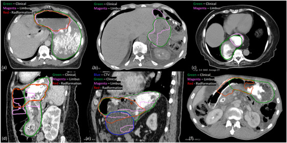
The autosegmentation software, developed by Limbus AI, MIM Contour ProtégéAI, Radformation AutoContour, and Siemens syngo.via, were used to segment 111 patient cases. Geometric segmentation accuracy was quantitatively compared with clinical contours using the dice similarity coefficient (DSC) and 95% Hausdorff distance (HD95). The outliers from quantitative evaluations of each software were analyzed for the liver, stomach, and kidneys with the possible causes of outliers summarized into six categories: (1) difference in contouring style or guideline, (2) image acquisition and quality, (3) abnormal anatomy of the OAR, (4) abnormal anatomy of abutting organs/tissues, (5) external/internal devices, and (6) other causes.

RESULTS

For the liver segmentation, the most prominent cause of discrepancies for Limbus, which occurred in four of its six outliers, was the existence of biliary stent or internal/external biliary drain as well as the resulting pneumobilia. Siemens included the abutting organs that shared CT numbers similar to those of the liver in 5/8 outliers. 12 of 13 Radformation's liver segmentation outliers included the heart and/or stomach while MIM not only included the stomach in the presence of barium in 5/11 outliers, but also produced fragmented contours in 5/11 other cases. Only Limbus and Radformation provided stomach segmentation, and imaging with barium contrast directly caused incomplete stomach delineation in 10/12 Limbus outliers and 21/25 Radformation outliers. As for the kidneys, Radformation and Siemens consistently followed the RTOG contouring guidelines, whereas the institutional contours excluded the renal pelvis in some cases, resulting in 19/25 Radformation outliers and 18/23 Siemens outliers. By contrast, Limbus contours appeared to follow different contouring guidelines that exclude the renal pelvis. Fragmented kidney contours were found in 10/15 Limbus outliers and 25/26 MIM outliers. The ones in MIM were directly linked to the use of IV contrast in imaging, but there was not enough evidence to identify the origin of Limbus's fragmented contours.

CONCLUSION

The causes of the segmentation outliers of the four commercial deep learning-based autocontouring solutions were summarized for each OAR. This work can help the vendors improve their autosegmentation software and also inform the users of potential modes of failure when using the tools.

摘要

目的

基于深度学习的危及器官(OAR)分割正逐渐成为临床实践的主流,因为其性能优于基于图谱和模型的自动轮廓描绘方法。虽然现在有几种基于深度学习的商业自动分割解决方案,但这些工具的实施仍处于非常原始的阶段,由于缺乏对系统分割倾向和失败模式的了解,验收标准尚不完善。作为临床实施迭代过程的起点,本研究聚焦于对四种用于腹部OAR的商业自动轮廓描绘工具的异常值分析。

材料与方法

使用由Limbus AI、MIM Contour ProtégéAI、Radformation AutoContour和西门子syngo.via开发的自动分割软件对111例患者病例进行分割。使用骰子相似系数(DSC)和95%豪斯多夫距离(HD95)将几何分割精度与临床轮廓进行定量比较。对每个软件定量评估中的异常值进行肝脏、胃和肾脏的分析,并将异常值的可能原因归纳为六类:(1)轮廓描绘风格或指南的差异,(2)图像采集与质量,(3)OAR的解剖结构异常,(4)相邻器官/组织的解剖结构异常,(5)外部/内部装置,以及(6)其他原因。

结果

对于肝脏分割,Limbus在其六个异常值中有四个出现差异的最主要原因是存在胆管支架或内部/外部胆管引流以及由此产生的气腹。西门子在其八个异常值中有五个将与肝脏CT值相似的相邻器官包括在内。Radformation的13个肝脏分割异常值中有12个包括心脏和/或胃,而MIM不仅在11个异常值中有5个在存在钡剂时将胃包括在内,而且在另外11个病例中有5个产生了破碎的轮廓。只有Limbus和Radformation提供胃分割,钡剂造影成像直接导致12个Limbus异常值中有10个胃轮廓描绘不完整,25个Radformation异常值中有21个胃轮廓描绘不完整。至于肾脏,Radformation和西门子始终遵循RTOG轮廓描绘指南,而机构轮廓在某些情况下排除了肾盂,导致25个Radformation异常值中有19个,23个西门子异常值中有18个。相比之下,Limbus轮廓似乎遵循不同的轮廓描绘指南,排除了肾盂。在15个Limbus异常值中有10个发现肾脏轮廓破碎,26个MIM异常值中有25个发现肾脏轮廓破碎。MIM中的破碎轮廓与成像中使用静脉造影剂直接相关,但没有足够证据确定Limbus破碎轮廓的来源。

结论

针对每种OAR总结了四种基于深度学习的商业自动轮廓描绘解决方案的分割异常值原因。这项工作有助于供应商改进其自动分割软件,也能让用户了解使用这些工具时潜在的失败模式。

https://cdn.ncbi.nlm.nih.gov/pmc/blobs/596b/11969109/920e8a2b5156/ACM2-26-e70010-g002.jpg
https://cdn.ncbi.nlm.nih.gov/pmc/blobs/596b/11969109/ade78d2d789c/ACM2-26-e70010-g004.jpg
https://cdn.ncbi.nlm.nih.gov/pmc/blobs/596b/11969109/e03619bd5463/ACM2-26-e70010-g001.jpg
https://cdn.ncbi.nlm.nih.gov/pmc/blobs/596b/11969109/a7b75150e47d/ACM2-26-e70010-g003.jpg
https://cdn.ncbi.nlm.nih.gov/pmc/blobs/596b/11969109/920e8a2b5156/ACM2-26-e70010-g002.jpg

相似文献

1
Evaluation and failure analysis of four commercial deep learning-based autosegmentation software for abdominal organs at risk.四种基于深度学习的腹部危险器官自动分割商用软件的评估与故障分析
J Appl Clin Med Phys. 2025 Apr;26(4):e70010. doi: 10.1002/acm2.70010. Epub 2025 Feb 13.
2
Evaluation of multiple-vendor AI autocontouring solutions.多厂商 AI 自动勾画解决方案评估。
Radiat Oncol. 2024 May 31;19(1):69. doi: 10.1186/s13014-024-02451-4.
3
Evaluation of deep learning-based autosegmentation in breast cancer radiotherapy.深度学习自动分割在乳腺癌放疗中的评价。
Radiat Oncol. 2021 Oct 14;16(1):203. doi: 10.1186/s13014-021-01923-1.
4
Investigation on performance of multiple AI-based auto-contouring systems in organs at risks (OARs) delineation.多个人工智能自动勾画系统在危险器官(OARs)勾画中的性能研究。
Phys Eng Sci Med. 2024 Sep;47(3):1123-1140. doi: 10.1007/s13246-024-01434-9. Epub 2024 Sep 2.
5
Evaluation of AI-based auto-contouring tools in radiotherapy: A single-institution study.基于人工智能的放疗自动轮廓勾画工具的评估:一项单机构研究。
J Appl Clin Med Phys. 2025 Apr;26(4):e14620. doi: 10.1002/acm2.14620. Epub 2025 Jan 21.
6
Geometric evaluations of CT and MRI based deep learning segmentation for brain OARs in radiotherapy.基于 CT 和 MRI 的深度学习分割的脑 OAR 几何评估在放射治疗中的应用。
Phys Med Biol. 2023 Aug 29;68(17). doi: 10.1088/1361-6560/acf023.
7
Gross failure rates and failure modes for a commercial AI-based auto-segmentation algorithm in head and neck cancer patients.商业人工智能自动分割算法对头颈部癌症患者的总体失败率和失败模式。
J Appl Clin Med Phys. 2024 Jun;25(6):e14273. doi: 10.1002/acm2.14273. Epub 2024 Jan 23.
8
Evaluation of a deep image-to-image network (DI2IN) auto-segmentation algorithm across a network of cancer centers.在一个癌症中心网络中对深度图像到图像网络(DI2IN)自动分割算法进行评估。
J Cancer Res Ther. 2024 Apr 1;20(3):1020-1025. doi: 10.4103/jcrt.jcrt_769_23. Epub 2024 Jan 22.
9
Dosimetric impact of contour editing on CT and MRI deep-learning autosegmentation for brain OARs.CT 和 MRI 深度学习自动勾画脑 OAR 时轮廓编辑对剂量学的影响。
J Appl Clin Med Phys. 2024 May;25(5):e14345. doi: 10.1002/acm2.14345. Epub 2024 Apr 25.
10
Comparative clinical evaluation of atlas and deep-learning-based auto-segmentation of organ structures in liver cancer.基于 atlas 和深度学习的肝癌器官结构自动分割的临床对比评估。
Radiat Oncol. 2019 Nov 27;14(1):213. doi: 10.1186/s13014-019-1392-z.

引用本文的文献

1
Can Deep Learning-Based Auto-Contouring Software Achieve Accurate Pelvic Volume Delineation in Volumetric Image-Guided Radiotherapy for Prostate Cancer? A Preliminary Multicentric Analysis.基于深度学习的自动轮廓勾画软件在前列腺癌容积图像引导放疗中能否实现准确的盆腔体积勾画?一项初步多中心分析。
Curr Oncol. 2025 May 30;32(6):321. doi: 10.3390/curroncol32060321.

本文引用的文献

1
Multicenter Study of Pelvic Nodal Autosegmentation Algorithm of Siemens Healthineers: Comparison of Male Versus Female Pelvis.西门子医疗集团盆腔淋巴结自动分割算法的多中心研究:男性与女性骨盆的比较
Adv Radiat Oncol. 2023 Jul 28;9(2):101326. doi: 10.1016/j.adro.2023.101326. eCollection 2024 Feb.
2
Clinical Implementation and Evaluation of Auto-Segmentation Tools for Multi-Site Contouring in Radiotherapy.放射治疗中多部位轮廓自动分割工具的临床应用与评估
Phys Imaging Radiat Oncol. 2023 Nov 17;28:100515. doi: 10.1016/j.phro.2023.100515. eCollection 2023 Oct.
3
NRG Oncology Assessment of Artificial Intelligence Deep Learning-Based Auto-segmentation for Radiation Therapy: Current Developments, Clinical Considerations, and Future Directions.
NRG 肿瘤学评估人工智能深度学习的自动分割在放射治疗中的应用:当前进展、临床考虑因素和未来方向。
Int J Radiat Oncol Biol Phys. 2024 May 1;119(1):261-280. doi: 10.1016/j.ijrobp.2023.10.033. Epub 2023 Nov 14.
4
Framework for Radiation Oncology Department-wide Evaluation and Implementation of Commercial Artificial Intelligence Autocontouring.放射肿瘤学部门范围内评估和实施商业人工智能自动勾画的框架。
Pract Radiat Oncol. 2024 Mar-Apr;14(2):e150-e158. doi: 10.1016/j.prro.2023.10.011. Epub 2023 Nov 5.
5
A clinical evaluation of the performance of five commercial artificial intelligence contouring systems for radiotherapy.五种商用人工智能放疗轮廓勾画系统性能的临床评估
Front Oncol. 2023 Aug 4;13:1213068. doi: 10.3389/fonc.2023.1213068. eCollection 2023.
6
Incremental retraining, clinical implementation, and acceptance rate of deep learning auto-segmentation for male pelvis in a multiuser environment.深度学习自动分割在多用户环境下对男性骨盆的增量再训练、临床实施和接受率。
Med Phys. 2023 Jul;50(7):4079-4091. doi: 10.1002/mp.16537. Epub 2023 Jun 7.
7
How smart is artificial intelligence in organs delineation? Testing a CE and FDA-approved Deep-Learning tool using multiple expert contours delineated on planning CT images.人工智能在器官轮廓描绘方面有多智能?使用在计划CT图像上描绘的多个专家轮廓测试一种获得CE和FDA批准的深度学习工具。
Front Oncol. 2023 Mar 2;13:1089807. doi: 10.3389/fonc.2023.1089807. eCollection 2023.
8
Implementation of a Commercial Deep Learning-Based Auto Segmentation Software in Radiotherapy: Evaluation of Effectiveness and Impact on Workflow.基于深度学习的商业自动分割软件在放射治疗中的应用:有效性评估及对工作流程的影响
Life (Basel). 2022 Dec 13;12(12):2088. doi: 10.3390/life12122088.
9
A clinical and time savings evaluation of a deep learning automatic contouring algorithm.一种深度学习自动轮廓算法的临床及节省时间评估
Med Dosim. 2023;48(1):55-60. doi: 10.1016/j.meddos.2022.11.001. Epub 2022 Dec 20.
10
Quality Assurance for AI-Based Applications in Radiation Therapy.放射治疗中基于人工智能的应用的质量保证。
Semin Radiat Oncol. 2022 Oct;32(4):421-431. doi: 10.1016/j.semradonc.2022.06.011.