Soni Swati, Walke Vaishali, Asati Dinesh, Maurya Anand, Mukhopadhyay Sramana
Peoples College of Medical Sciences and Research Centre, Bhopal (MP), India.
Path & Lab Medicine, All India Institute of Medical Science, Bhopal. India.
Iran J Pathol. 2025;20(1):33-41. doi: 10.30699/ijp.2024.2023590.3263. Epub 2025 Jan 10.
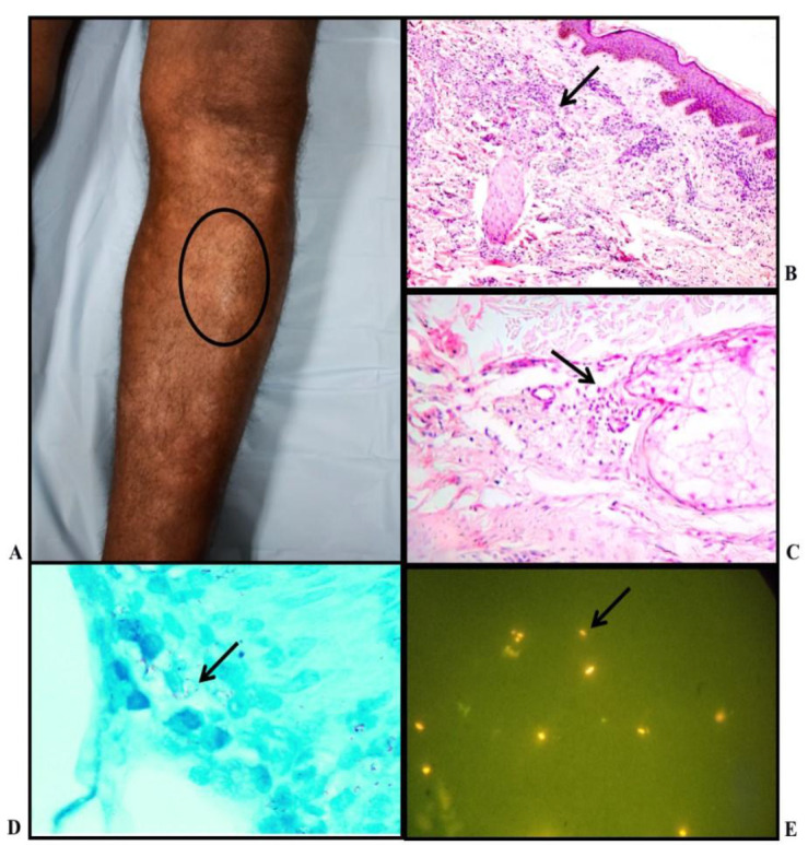
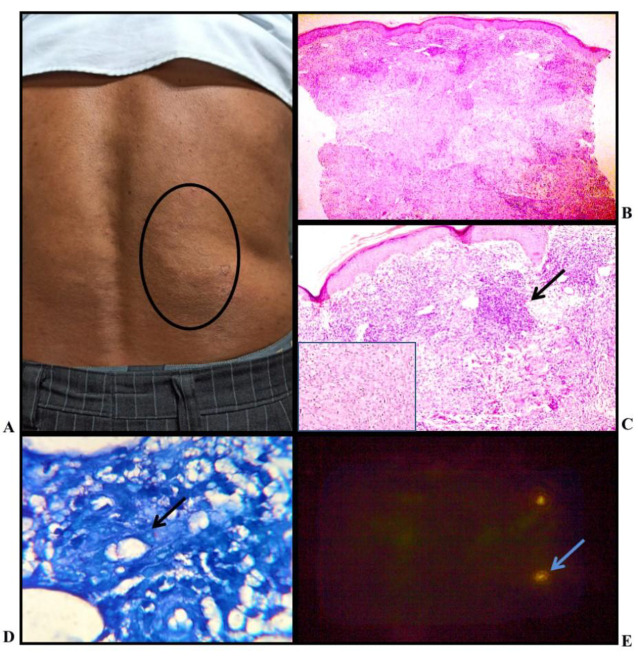
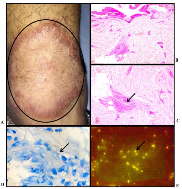
BACKGROUND & OBJECTIVE: Leprosy is a chronic infectious disease caused by Mycobacterium leprae. Fite-Faraco (FF) is the routine staining method used to demonstrate the presence of in tissue sections. Fluorescent microscopy (FM) can help visualize lepra bacilli better. The present study compares two methodologies, fluorescent microscopy, and Fite-Faraco, in detecting Mycobacterium leprae in tissue sections.
Histopathology of skin biopsies in 60 cases of Hansen's were evaluated with FF stain. The performance of Auramine- Rhodamine Fluroscencent stain was compared with conventional FF staining in identifying Lepra bacilli.
A total of 60 clinically and histopathologically confirmed cases of Hansen's disease were included in this ambispective study. The cases were sub-classified into various histological categories. Auramine-rhodamine fluorescent staining was performed and examined under a fluorescent microscope with an LED light illuminator. The bacteriological index (BI) was calculated under an oil immersion field for both Fite-Faraco (FF) staining and fluorescent microscopy (FM), graded from zero to six plus according to Ridley's logarithmic scale. Lepra bacilli were identified in 70% of patients on FF staining, while fluorescent microscopy showed positivity in 80%. The mean BI calculated by FM (2.48) was significantly higher than that by the FF method (2.18), and more multibacillary disease was identified by fluorescent staining compared to FF staining.
It is advantageous to use fluorescent microscopy as an adjunct to conventional Fite-Faraco stain especially in cases where the latter fails to detect lepra bacilli and in a clinically suspected multibacillary disease.
麻风病是一种由麻风分枝杆菌引起的慢性传染病。菲-法拉科(Fite-Faraco,FF)染色法是用于在组织切片中显示麻风杆菌的常规染色方法。荧光显微镜检查(FM)有助于更好地观察麻风杆菌。本研究比较了荧光显微镜检查和菲-法拉科染色法这两种方法在检测组织切片中麻风分枝杆菌方面的效果。
对60例麻风病患者的皮肤活检组织进行组织病理学检查,采用FF染色。将金胺-罗丹明荧光染色法与传统的FF染色法在鉴定麻风杆菌方面的性能进行比较。
本双盲前瞻性研究共纳入60例临床和组织病理学确诊的麻风病病例。这些病例被分为不同的组织学类别。进行金胺-罗丹明荧光染色,并在配备LED光源的荧光显微镜下检查。分别根据菲-法拉科(FF)染色法和荧光显微镜检查(FM)在油镜视野下计算细菌指数(BI),按照里德利对数分级标准从0到6+进行分级。FF染色法在70%的患者中检测到麻风杆菌,而荧光显微镜检查显示阳性率为80%。FM计算得出的平均BI(2.48)显著高于FF法(2.18),与FF染色相比,荧光染色法检测出更多的多菌型病例。
尤其在传统的菲-法拉科染色法未能检测到麻风杆菌的病例以及临床疑似多菌型疾病的情况下,将荧光显微镜检查作为传统菲-法拉科染色的辅助方法具有优势。