Kim Donghee, Jung Ji Seung, Hwang Jiyi, Park Jiwoo, Kwon Myeongjee, Yong Jungyeon, Yoon Haerin, Park Kyung-Mee
Laboratory of Veterinary Surgery and Ophthalmology, College of Veterinary Medicine, Chungbuk National University, Cheongju, Korea.
BMC Vet Res. 2025 Mar 19;21(1):181. doi: 10.1186/s12917-025-04648-5.
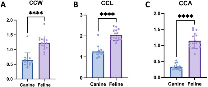
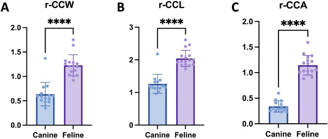
This study aims to investigate the anatomical differences in the anterior segment of the eyes between dogs and cats using ultrasound biomicroscopy (UBM) to understand the higher prevalence of primary angle-closure glaucoma (PACG) in dogs compared to cats. Retrospective analysis was performed on clinical data from 16 eyes of 16 dogs and 14 eyes of 14 cats with normal eye conditions. UBM was utilized to measure nine specific parameters, including Schwalbe's Line Distance (SLD), Iridocorneal Angle (ICA), Angle-Opening Distance (AOD), and three ciliary cleft parameters: width (CCW), length (CCL), and area (CCA). To account for differences in body size, ciliary cleft parameters were adjusted accordingly.
Significant anatomical differences in the anterior segment were found between the two species. Dogs had smaller values for SLD, ICA, AOD, and ciliary cleft parameters (CCW, CCL, CCA) compared to cats. Even after body-size adjustment, the rectified ciliary cleft parameters remained smaller in dogs.
The anatomical differences, particularly the smaller ciliary cleft and narrower drainage angles in dogs, may contribute to the higher prevalence of PACG in this species. Conversely, the larger ciliary cleft in cats may explain the lower occurrence of primary glaucoma in cats.
本研究旨在利用超声生物显微镜(UBM)研究犬猫眼前段的解剖差异,以了解犬原发性闭角型青光眼(PACG)患病率高于猫的原因。对16只犬的16只眼和14只猫的14只眼的正常眼部临床数据进行回顾性分析。使用UBM测量九个特定参数,包括施瓦贝线距离(SLD)、虹膜角膜角(ICA)、房角开放距离(AOD)以及三个睫状体间隙参数:宽度(CCW)、长度(CCL)和面积(CCA)。为了考虑体型差异,相应地对睫状体间隙参数进行了调整。
发现两个物种的眼前段存在显著的解剖差异。与猫相比,犬的SLD、ICA、AOD和睫状体间隙参数(CCW、CCL、CCA)值较小。即使在调整体型后,犬的校正睫状体间隙参数仍然较小。
解剖差异,特别是犬的睫状体间隙较小和引流角较窄,可能是该物种PACG患病率较高的原因。相反,猫的睫状体间隙较大可能解释了猫原发性青光眼发生率较低的原因。