Papadopoulos Petros, Zisis Vasileios, Andreadis Dimitrios, Poulopoulos Konstantinos, Parlitsis Dimitrios, Paraskevopoulos Konstantinos, Anastasiadou Pinelopi A, Anagnostou Eleftherios, Vahtsevanos Konstantinos, Poulopoulos Athanasios
Oral Medicine/ Pathology, Aristotle University of Thessaloniki, Thessaloniki, GRC.
Oral and Maxillofacial Surgery, Papanikolaou Hospital, Aristotle University of Thessaloniki, Thessaloniki, GRC.
Cureus. 2025 Feb 16;17(2):e79085. doi: 10.7759/cureus.79085. eCollection 2025 Feb.
The silencing of death-associated protein kinase 1 (DAPK-1) is an effective way of inactivating a tumor-suppressing mechanism. The aim of this study was to investigate the immunohistochemical expression of DAPK-1 in oral leukoplakia (OL) and oral squamous cell carcinoma (OSCC).
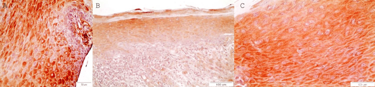
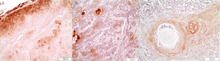
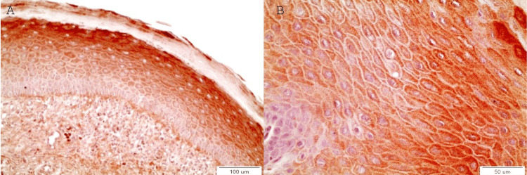
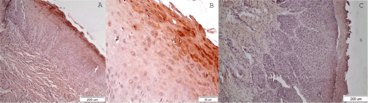
The immunohistochemical (IHC) detection of DAPK-1 was carried out in cases of OLs and OSCCs. DAPK-1 molecules' tissue distribution in OLs/OSCCs tissues was evaluated using semiquantitative immunohistochemistry in representative paraffin-embedded tissue samples (57 in total) from 2004-2019, retrieved from the archives of the Department of Oral Medicine/Pathology, School of Dentistry, Aristotle University of Thessaloniki, Greece and the St Lukas Hospital of Thessaloniki, Greece. The inclusion criterion was the presence of sufficient precancerous or cancerous biological material (estimated as more than 70% per tissue specimen) in the paraffin cubes. The exclusion criterion was the opposite, i.e. the lack of sufficient material due to previous sections. Statistics for IHC were evaluated by a non-parametric Mann-Whitney U Test. A two-sided p-value < 0.05 was considered statistically significant.
DAPK-1 IHC expression was increased in OLs without dysplasia and with OLs with mild dysplasia compared to moderate/severe dysplasia (p=0.019, Mann-Whitney U Test) and OSCCs (p=0.003, Mann-Whitney U Test). Conclusions: DAPK-1 seemed to function as an oncosuppressor molecular biomarker, as its expression was decreased in areas of cellular dysplasia in OLs and in areas of OSCCs composed of less differentiated cells. The clinical application of this biomarker is that the positively stained, potentially malignant lesions are less likely to transition into malignancy, and cancerous lesions are more likely to behave non-aggressively. On the other hand, the lack of staining could signify the loss of this oncosuppressing ability, and it could be a potential prognostic biomarker for OSCC's aggressive biologic behavior if considered with other clinical parameters and a prognostic factor of malignant transformation of potentially malignant lesions. Since this is a preliminary study, more studies with larger sample sizes are required to support these conclusions.
死亡相关蛋白激酶1(DAPK-1)的沉默是使肿瘤抑制机制失活的一种有效方式。本研究的目的是调查DAPK-1在口腔白斑(OL)和口腔鳞状细胞癌(OSCC)中的免疫组化表达情况。
对OL和OSCC病例进行DAPK-1的免疫组化(IHC)检测。使用半定量免疫组化方法评估DAPK-1分子在OL/OSCC组织中的组织分布情况,这些组织样本来自2004年至2019年希腊塞萨洛尼基亚里士多德大学牙科学院口腔医学/病理学系档案以及希腊塞萨洛尼基圣卢卡斯医院,共57个代表性石蜡包埋组织样本。纳入标准是石蜡块中存在足够的癌前或癌性生物材料(每个组织标本估计超过70%)。排除标准则相反,即由于先前切片导致缺乏足够的材料。通过非参数曼-惠特尼U检验评估IHC的统计学结果。双侧p值<0.05被认为具有统计学意义。
与中度/重度发育异常的OL和OSCC相比,DAPK-1的IHC表达在无发育异常的OL和轻度发育异常的OL中有所增加(曼-惠特尼U检验,p = 0.019)以及在OSCC中(曼-惠特尼U检验,p = 0.003)。结论:DAPK-1似乎作为一种肿瘤抑制分子生物标志物发挥作用,因为其在OL的细胞发育异常区域以及由分化程度较低的细胞组成的OSCC区域中表达降低。这种生物标志物的临床应用在于,阳性染色的潜在恶性病变转变为恶性的可能性较小,而癌性病变的侵袭性可能较低。另一方面,缺乏染色可能表明这种肿瘤抑制能力的丧失,如果与其他临床参数一起考虑,它可能是OSCC侵袭性生物学行为的潜在预后生物标志物,也是潜在恶性病变恶性转化的预后因素。由于这是一项初步研究,需要更多样本量更大的研究来支持这些结论。