Greene Whitney, Pereira Nuno, Doescher Bethany, Rojo-Solis Carlos, David Hugo, Faustino Ricardo, Reese David, De Voe Ryan, Latson Ed, Mylniczenko Natalie
Mote Marine Laboratory and Aquarium, Sarasota, FL, United States.
Oceanário de Lisboa, Esplanada D. Carlos I, Lisbon, Portugal.
Front Vet Sci. 2025 Mar 10;11:1463428. doi: 10.3389/fvets.2024.1463428. eCollection 2024.
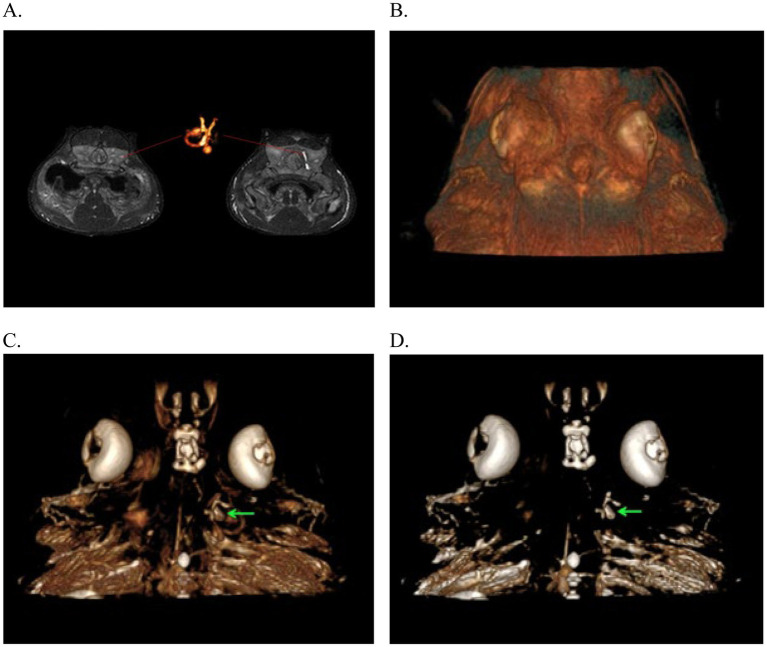
The inner ear is an often overlooked system in elasmobranchs with few documented reports of disease or other abnormalities in the literature. Similar to terrestrial vertebrates, it is located in the cranium, and there are multiple components to the ear of elasmobranchs including a pair of membranous labyrinths each with three semicircular canals and four chambers or end organs (the saccule, the lagena, the utricle and the macula neglecta) making up the endolymphatic system (ELS). There is species variability among the inner ear anatomy of elasmobranchs, and this may play a role in disease development, progression, and treatment outcomes. Also similar to terrestrial vertebrates, this system plays a key role in hearing, acceleration, and orientation. When affected, clinical signs may include localized areas of swelling or stoma development along the dorsal midline of the head at the endolymphatic pores, atypical swimming behaviors consistent with vestibular disease (spiraling/spinning or barrel rolling, or tilting to one side), and anorexia. Less frequently, the eyes may also be affected and present with exophthalmia, hyphema, and/or panophthalmitis. Herein are case series from five institutions representing a variety of elasmobranch species affected with ELS disease with discussion of anatomy, clinical presentation, diagnostics, etiology, treatment, and outcomes. Endolymphatic disease may be clinically underdiagnosed in elasmobranchs and mistaken for other diseases such as superficial subcutaneous or subdermal abscesses, focal dermatitis, or neuropathies presumed to not be associated with the inner ear system. In addition, disease may be occult for a long period of time prior to overt manifestation of signs or chronic with waxing and waning clinical signs, likely because of anatomy and resultant treatment challenges. Awareness and additional research may help to promote timely identification, improve diagnostic and treatment options, and help to optimize individual animal welfare.
内耳是板鳃亚纲动物中一个常被忽视的系统,文献中关于该系统疾病或其他异常的记载报道较少。与陆生脊椎动物类似,内耳位于颅骨内,板鳃亚纲动物的耳朵有多个组成部分,包括一对膜迷路,每个膜迷路有三个半规管和四个腔室或终器(球囊、瓶状囊、椭圆囊和忽视斑),共同构成内淋巴系统(ELS)。板鳃亚纲动物内耳的解剖结构存在物种差异,这可能在疾病的发生、发展和治疗结果中发挥作用。同样与陆生脊椎动物相似,该系统在听觉、加速和定向方面起着关键作用。当受到影响时,临床症状可能包括内淋巴孔处头部背中线沿线局部肿胀或气孔形成区域、与前庭疾病一致的非典型游泳行为(螺旋式/旋转或翻滚,或向一侧倾斜)以及厌食。较少见的情况下,眼睛也可能受到影响,出现眼球突出、前房积血和/或全眼球炎。本文介绍了来自五个机构的病例系列,这些病例涉及多种受ELS疾病影响的板鳃亚纲动物物种,并讨论了解剖结构、临床表现、诊断、病因、治疗和结果。内淋巴疾病在板鳃亚纲动物中可能在临床上被漏诊,并被误诊为其他疾病,如浅表皮下或皮内脓肿、局灶性皮炎或推测与内耳系统无关的神经病变。此外,在症状明显出现之前,疾病可能长期隐匿,或者具有慢性且临床症状时好时坏,这可能是由于解剖结构以及由此带来的治疗挑战所致。提高认识和开展更多研究可能有助于促进及时识别、改善诊断和治疗选择,并有助于优化个体动物的福利。