Provenzano Destie, Wang Jeffrey, Goyal Sharad, Rao Yuan James
School of Engineering and Applied Science, George Washington University, Washington, DC 20052, USA.
Department of Radiation Oncology, School of Medicine and Health Sciences, George Washington University, Washington, DC 20052, USA.
Tomography. 2025 Mar 20;11(3):38. doi: 10.3390/tomography11030038.
Predictive models like Residual Neural Networks (ResNets) can use Magnetic Resonance Imaging (MRI) data to identify cervix tumors likely to recur after radiotherapy (RT) with high accuracy. However, there persists a lack of insight into model selections (explainability). In this study, we explored whether model features could be used to generate simulated images as a method of model explainability.
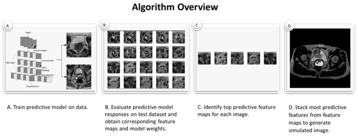
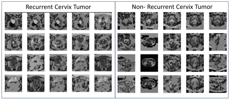
T2W MRI data were collected for twenty-seven women with cervix cancer who received RT from the TCGA-CESC database. Simulated images were generated as follows: [A] a ResNet model was trained to identify recurrent cervix cancer; [B] a model was evaluated on T2W MRI data for subjects to obtain corresponding feature maps; [C] most important feature maps were determined for each image; [D] feature maps were combined across all images to generate a simulated image; [E] the final image was reviewed by a radiation oncologist and an initial algorithm to identify the likelihood of recurrence.
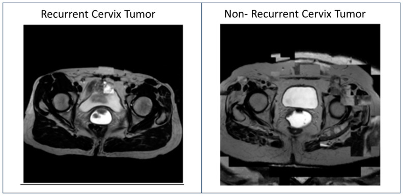
Predictive feature maps from the ResNet model (93% accuracy) were used to generate simulated images. Simulated images passed through the model were identified as recurrent and non-recurrent cervix tumors after radiotherapy. A radiation oncologist identified the simulated images as cervix tumors with characteristics of aggressive Cervical Cancer. These images also contained multiple MRI features not considered clinically relevant.
This simple method was able to generate simulated MRI data that mimicked recurrent and non-recurrent cervix cancer tumor images. These generated images could be useful for evaluating the explainability of predictive models and to assist radiologists with the identification of features likely to predict disease course.
像残差神经网络(ResNets)这样的预测模型可以利用磁共振成像(MRI)数据高精度地识别放疗(RT)后可能复发的子宫颈肿瘤。然而,对于模型选择(可解释性)仍缺乏深入了解。在本研究中,我们探讨了是否可以使用模型特征来生成模拟图像作为一种模型可解释性的方法。
从TCGA - CESC数据库收集了27名接受放疗的子宫颈癌女性的T2W MRI数据。模拟图像的生成如下:[A]训练一个ResNet模型以识别复发性子宫颈癌;[B]在受试者的T2W MRI数据上评估一个模型以获得相应的特征图;[C]为每个图像确定最重要的特征图;[D]将所有图像的特征图组合以生成模拟图像;[E]由放射肿瘤学家和初始算法审查最终图像以识别复发的可能性。
使用来自ResNet模型的预测特征图(准确率93%)生成模拟图像。通过该模型的模拟图像在放疗后被识别为复发性和非复发性子宫颈肿瘤。一名放射肿瘤学家将模拟图像识别为具有侵袭性子宫颈癌特征的子宫颈肿瘤。这些图像还包含多个临床上不被认为相关的MRI特征。
这种简单方法能够生成模拟MRI数据,模拟复发性和非复发性子宫颈癌肿瘤图像。这些生成的图像可能有助于评估预测模型的可解释性,并协助放射科医生识别可能预测疾病进程的特征。