• 文献检索
  • 文档翻译
  • 深度研究
  • 学术资讯
  • Suppr Zotero 插件Zotero 插件
  • 邀请有礼
  • 套餐&价格
  • 历史记录
应用&插件
Suppr Zotero 插件Zotero 插件浏览器插件Mac 客户端Windows 客户端微信小程序
定价
高级版会员购买积分包购买API积分包
服务
文献检索文档翻译深度研究API 文档MCP 服务
关于我们
关于 Suppr公司介绍联系我们用户协议隐私条款
关注我们

Suppr 超能文献

核心技术专利:CN118964589B侵权必究
粤ICP备2023148730 号-1Suppr @ 2026

文献检索

告别复杂PubMed语法,用中文像聊天一样搜索,搜遍4000万医学文献。AI智能推荐,让科研检索更轻松。

立即免费搜索

文件翻译

保留排版,准确专业,支持PDF/Word/PPT等文件格式,支持 12+语言互译。

免费翻译文档

深度研究

AI帮你快速写综述,25分钟生成高质量综述,智能提取关键信息,辅助科研写作。

立即免费体验

使用深度学习对光学相干断层扫描血管内成像中的冠状动脉斑块进行自动综合评估。

Automated comprehensive evaluation of coronary artery plaque in IVOCT using deep learning.

作者信息

Liu Pengfei, Lu Zang, Hou Wenqing, Kadier Kaisaierjiang, Cui Chunying, Mu Zhengyang, Ainiwaer Aikeliyaer, Peng Xinliang, Wufu Gulinuer, Ma Yitong, Dai Jianguo, Ma Xiang

机构信息

Department of Cardiology, First Affiliated Hospital of Xinjiang Medical University, Urumqi, China.

College of Information Science and Technology, Shihezi University, Shihezi 832003, Xinjiang, China.

出版信息

iScience. 2025 Mar 6;28(4):112169. doi: 10.1016/j.isci.2025.112169. eCollection 2025 Apr 18.

DOI:10.1016/j.isci.2025.112169
PMID:40224006
原文链接:https://pmc.ncbi.nlm.nih.gov/articles/PMC11987667/
Abstract

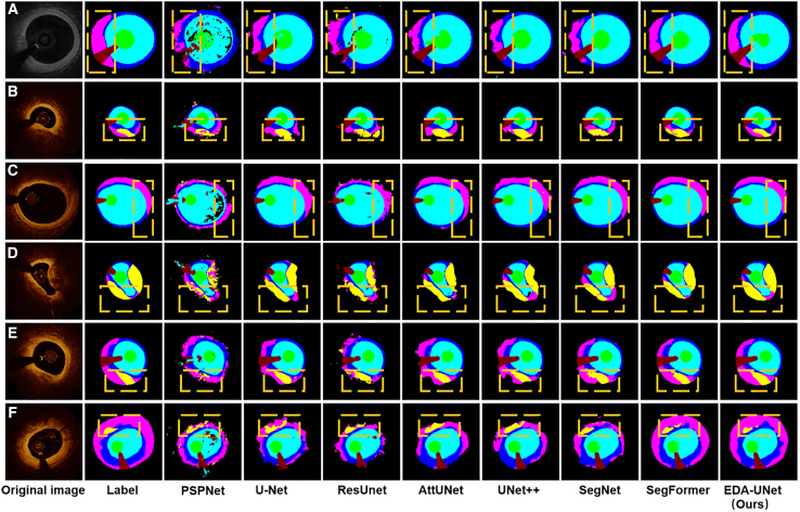
The process of manually characterizing and quantifying coronary artery plaque tissue in intravascular optical coherence tomography (IVOCT) images is both time-consuming and subjective. We have developed a deep learning-based semantic segmentation model (EDA-UNet) designed specifically for characterizing and quantifying coronary artery plaque tissue in IVOCT images. IVOCT images from two centers were utilized as the internal dataset for model training and internal testing. Images from another independent center employing IVOCT were used for external testing. The Dice coefficients for fibrous plaque, calcified plaque, and lipid plaque in external tests were 0.8282, 0.7408, and 0.7052 respectively. The model demonstrated strong correlation and consistency with the ground truth in the quantitative analysis of calcification scores and the identification of thin-cap fibroatheroma (TCFA). The median duration for each callback analysis was 18 s. EDA-UNet model serves as an efficient and accurate technological tool for plaque characterization and quantification.

摘要

在血管内光学相干断层扫描(IVOCT)图像中手动表征和量化冠状动脉斑块组织的过程既耗时又主观。我们开发了一种基于深度学习的语义分割模型(EDA-UNet),专门用于表征和量化IVOCT图像中的冠状动脉斑块组织。来自两个中心的IVOCT图像被用作模型训练和内部测试的内部数据集。来自另一个采用IVOCT的独立中心的图像用于外部测试。外部测试中纤维斑块、钙化斑块和脂质斑块的Dice系数分别为0.8282、0.7408和0.7052。该模型在钙化评分的定量分析和薄帽纤维粥样瘤(TCFA)的识别中与地面真值表现出很强的相关性和一致性。每次回调分析的中位持续时间为18秒。EDA-UNet模型是一种用于斑块表征和量化的高效、准确的技术工具。

https://cdn.ncbi.nlm.nih.gov/pmc/blobs/27d3/11987667/3b1c818df6dd/gr8.jpg
https://cdn.ncbi.nlm.nih.gov/pmc/blobs/27d3/11987667/cdd288cfe825/fx1.jpg
https://cdn.ncbi.nlm.nih.gov/pmc/blobs/27d3/11987667/2ad3c5f3b9d0/gr1.jpg
https://cdn.ncbi.nlm.nih.gov/pmc/blobs/27d3/11987667/8935355cd4ed/gr2.jpg
https://cdn.ncbi.nlm.nih.gov/pmc/blobs/27d3/11987667/78926a74ff86/gr3.jpg
https://cdn.ncbi.nlm.nih.gov/pmc/blobs/27d3/11987667/d0236c9249b2/gr4.jpg
https://cdn.ncbi.nlm.nih.gov/pmc/blobs/27d3/11987667/80891bedc7a6/gr5.jpg
https://cdn.ncbi.nlm.nih.gov/pmc/blobs/27d3/11987667/d324e7b93df4/gr6.jpg
https://cdn.ncbi.nlm.nih.gov/pmc/blobs/27d3/11987667/7b808f91f7fc/gr7.jpg
https://cdn.ncbi.nlm.nih.gov/pmc/blobs/27d3/11987667/3b1c818df6dd/gr8.jpg

相似文献

1
Automated comprehensive evaluation of coronary artery plaque in IVOCT using deep learning.使用深度学习对光学相干断层扫描血管内成像中的冠状动脉斑块进行自动综合评估。
iScience. 2025 Mar 6;28(4):112169. doi: 10.1016/j.isci.2025.112169. eCollection 2025 Apr 18.
2
Automated analysis of fibrous cap in intravascular optical coherence tomography images of coronary arteries.冠状动脉血管内光学相干断层成像图像中纤维帽的自动分析。
Sci Rep. 2022 Dec 12;12(1):21454. doi: 10.1038/s41598-022-24884-1.
3
Deep learning segmentation of fibrous cap in intravascular optical coherence tomography images.基于血管内光学相干断层成像图像的深度学习纤维帽分割。
Sci Rep. 2024 Feb 22;14(1):4393. doi: 10.1038/s41598-024-55120-7.
4
Histology-Grounded Automated Plaque Subtype Segmentation in Intravascular Optical Coherence Tomography.基于组织学的血管内光学相干断层扫描斑块亚型自动分割
J Soc Cardiovasc Angiogr Interv. 2025 Mar 18;4(3Part B):102524. doi: 10.1016/j.jscai.2024.102524. eCollection 2025 Mar.
5
Diagnosis of Thin-Capped Fibroatheromas in Intravascular Optical Coherence Tomography Images: Effects of Light Scattering.血管内光学相干断层扫描图像中薄帽纤维粥样斑块的诊断:光散射的影响
Circ Cardiovasc Interv. 2016 Jul;9(7). doi: 10.1161/CIRCINTERVENTIONS.115.003163.
6
Artificial intelligence and optical coherence tomography for the automatic characterisation of human atherosclerotic plaques.人工智能和光学相干断层扫描技术在人类动脉粥样硬化斑块自动特征化中的应用。
EuroIntervention. 2021 May 17;17(1):41-50. doi: 10.4244/EIJ-D-20-01355.
7
OCTOPUS - Optical coherence tomography plaque and stent analysis software.章鱼 - 光学相干断层扫描斑块及支架分析软件。
Heliyon. 2023 Feb 1;9(2):e13396. doi: 10.1016/j.heliyon.2023.e13396. eCollection 2023 Feb.
8
Plaque burden can be assessed using intravascular optical coherence tomography and a dedicated automated processing algorithm: a comparison study with intravascular ultrasound.可以使用血管内光学相干断层扫描和专用的自动处理算法评估斑块负担:与血管内超声的对比研究。
Eur Heart J Cardiovasc Imaging. 2020 Jun 1;21(6):640-652. doi: 10.1093/ehjci/jez185.
9
Fibroatheroma identification in Intravascular Optical Coherence Tomography images using deep features.利用深度特征在血管内光学相干断层扫描图像中识别纤维粥样斑块。
Annu Int Conf IEEE Eng Med Biol Soc. 2017 Jul;2017:1501-1504. doi: 10.1109/EMBC.2017.8037120.
10
A-line-based thin-cap fibroatheroma detection with multi-view IVOCT images using multi-task learning and contrastive learning.基于 A 线的薄帽纤维粥样瘤多视图 IVOCT 图像检测:使用多任务学习和对比学习。
J Opt Soc Am A Opt Image Sci Vis. 2022 Dec 1;39(12):2298-2306. doi: 10.1364/JOSAA.464303.

本文引用的文献

1
Calcified coronary lesions: Imaging, prognosis, preparation and treatment state of the art review.钙化性冠状动脉病变:影像学、预后、准备和治疗的最新进展综述。
Prog Cardiovasc Dis. 2024 Sep-Oct;86:26-37. doi: 10.1016/j.pcad.2024.06.007. Epub 2024 Jun 24.
2
Artificial Intelligence for the Interventional Cardiologist: Powering and Enabling OCT Image Interpretation.介入心脏病专家的人工智能:助力并实现光学相干断层扫描(OCT)图像解读
Interv Cardiol. 2024 Mar 11;19:e03. doi: 10.15420/icr.2023.13. eCollection 2024.
3
Thin-cap fibroatheroma in acute coronary syndrome: Implication for intravascular imaging assessment.
急性冠脉综合征中的薄帽纤维粥样瘤:血管内影像学评估的意义。
Int J Cardiol. 2024 Jun 15;405:131965. doi: 10.1016/j.ijcard.2024.131965. Epub 2024 Mar 15.
4
A cascading learning method with SegFormer for radiographic measurement of periodontal bone loss.一种基于SegFormer的用于牙周骨丧失影像学测量的级联学习方法。
BMC Oral Health. 2024 Mar 11;24(1):325. doi: 10.1186/s12903-024-04079-y.
5
Role of intravascular ultrasound and optical coherence tomography in intracoronary imaging for coronary artery disease: a systematic review.血管内超声和光学相干断层扫描在冠状动脉疾病冠状动脉成像中的作用:一项系统评价。
J Geriatr Cardiol. 2024 Jan 28;21(1):104-129. doi: 10.26599/1671-5411.2024.01.001.
6
Deep learning segmentation of fibrous cap in intravascular optical coherence tomography images.基于血管内光学相干断层成像图像的深度学习纤维帽分割。
Sci Rep. 2024 Feb 22;14(1):4393. doi: 10.1038/s41598-024-55120-7.
7
A transformer-based pyramid network for coronary calcified plaque segmentation in intravascular optical coherence tomography images.基于变压器的金字塔网络用于血管内光学相干断层扫描图像中的冠状动脉钙化斑块分割。
Comput Med Imaging Graph. 2024 Apr;113:102347. doi: 10.1016/j.compmedimag.2024.102347. Epub 2024 Feb 9.
8
Reproducibility of an artificial intelligence optical coherence tomography software for tissue characterization: Implications for the design of longitudinal studies.人工智能光学相干断层扫描软件进行组织特征分析的可重复性:对纵向研究设计的影响。
Cardiovasc Revasc Med. 2024 Jan;58:79-87. doi: 10.1016/j.carrev.2023.07.003. Epub 2023 Jul 16.
9
Sk-Conv and SPP-based UNet for lesion segmentation of coronary optical coherence tomography.基于 Sk-Conv 和 SPP 的 UNet 用于冠状动脉光学相干断层扫描的病变分割。
Technol Health Care. 2023;31(S1):347-355. doi: 10.3233/THC-236030.
10
PDAtt-Unet: Pyramid Dual-Decoder Attention Unet for Covid-19 infection segmentation from CT-scans.PDAtt-Unet:用于从 CT 扫描中分割新冠感染的金字塔双解码器注意 U 型网络。
Med Image Anal. 2023 May;86:102797. doi: 10.1016/j.media.2023.102797. Epub 2023 Mar 21.