• 文献检索
  • 文档翻译
  • 深度研究
  • 学术资讯
  • Suppr Zotero 插件Zotero 插件
  • 邀请有礼
  • 套餐&价格
  • 历史记录
应用&插件
Suppr Zotero 插件Zotero 插件浏览器插件Mac 客户端Windows 客户端微信小程序
定价
高级版会员购买积分包购买API积分包
服务
文献检索文档翻译深度研究API 文档MCP 服务
关于我们
关于 Suppr公司介绍联系我们用户协议隐私条款
关注我们

Suppr 超能文献

核心技术专利:CN118964589B侵权必究
粤ICP备2023148730 号-1Suppr @ 2026

文献检索

告别复杂PubMed语法,用中文像聊天一样搜索,搜遍4000万医学文献。AI智能推荐,让科研检索更轻松。

立即免费搜索

文件翻译

保留排版,准确专业,支持PDF/Word/PPT等文件格式,支持 12+语言互译。

免费翻译文档

深度研究

AI帮你快速写综述,25分钟生成高质量综述,智能提取关键信息,辅助科研写作。

立即免费体验

通过具有CEM和超声整合的可解释机器学习模型提高乳腺癌腋窝淋巴结转移预测的特异性

Enhancing Specificity in Predicting Axillary Lymph Node Metastasis in Breast Cancer through an Interpretable Machine Learning Model with CEM and Ultrasound Integration.

作者信息

Xu Weimin, Zheng Bowen, Wen Chanjuan, Zeng Hui, Wang Sina, He Zilong, Liao Xin, Chen Weiguo, Li Yingjia, Qin Genggeng

机构信息

Department of Radiology, Nanfang Hospital, Southern Medical University, Guangzhou, China.

Department of Ultrasonic Medicine, NanfangHospital, Southern Medical University, Guangzhou, China.

出版信息

Technol Cancer Res Treat. 2025 Jan-Dec;24:15330338251334735. doi: 10.1177/15330338251334735. Epub 2025 Apr 17.

DOI:10.1177/15330338251334735
PMID:40241520
原文链接:https://pmc.ncbi.nlm.nih.gov/articles/PMC12035205/
Abstract

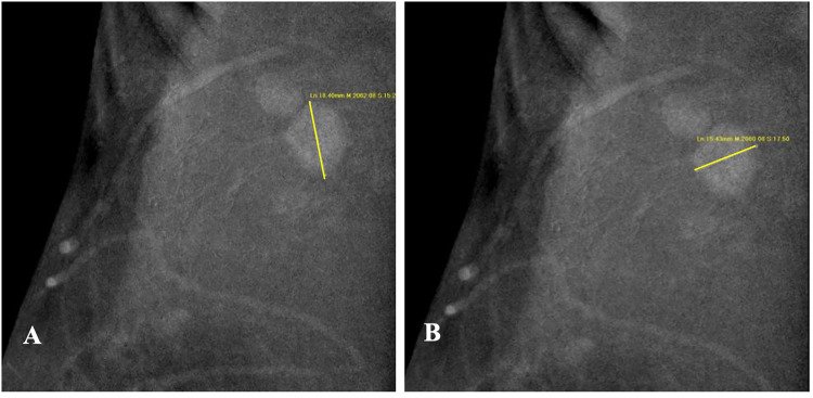
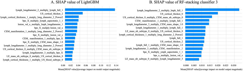
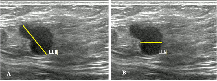
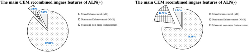
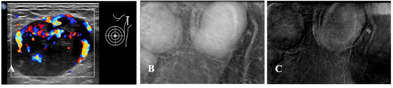
IntroductionThe study aims to evaluate the performance of an interpretable machine learning model in predicting preoperative axillary lymph node metastasis using primary breast cancer and lymph node features derived from contrast-enhanced mammography (CEM) and ultrasound (US) breast imaging reporting and data systems (BI-RADS).MethodsThis retrospective study included patients diagnosed with primary breast cancer. Two experienced radiologists extracted the BI-RADS features from the largest cross-section of the lesions and axillary lymph nodes based on CEM and US images, creating three datasets. Each dataset will train six base models to predict axillary lymph nodes, with pathological results serving as the gold standard. The top three models were used to train the five ensemble models. Additionally, SHapley Additive exPlanations (SHAP) was used to interpret the optimal model. The receiver-operating characteristic curve (ROC) and AUC were used to evaluate model performance.ResultsThis study involved 292 female patients, of whom 99 had axillary lymph node metastasis and 193 did not. The combination of CEM and ultrasound BI-RADS demonstrated the best performance in predicting axillary lymph node metastasis. Among these, the LightGBM achieved the highest AUC (0.762) and specificity (86.67%, while the ensemble model using RF as the meta-model had an AUC (0.754) and specificity (83.33%. The most important variables identified by SHAP were the long diameters of the lymph nodes in the CEM recombined image, along with their complete morphology in the low-energy image.ConclusionThe machine learning model using CEM and US BI-RADS features accurately predicted axillary lymph node metastasis before surgery, thereby serving as a valuable tool for clinical decision-making in patients with breast cancer.

摘要

引言

本研究旨在评估一种可解释的机器学习模型在利用原发性乳腺癌以及源自乳腺对比增强钼靶(CEM)和超声(US)乳腺影像报告和数据系统(BI-RADS)的淋巴结特征预测术前腋窝淋巴结转移方面的性能。

方法

这项回顾性研究纳入了被诊断为原发性乳腺癌的患者。两名经验丰富的放射科医生基于CEM和US图像从病变和腋窝淋巴结的最大横截面提取BI-RADS特征,创建了三个数据集。每个数据集将训练六个基础模型以预测腋窝淋巴结,病理结果作为金标准。使用排名前三的模型训练五个集成模型。此外,使用SHapley加性解释(SHAP)来解释最优模型。采用受试者操作特征曲线(ROC)和AUC评估模型性能。

结果

本研究涉及292名女性患者,其中99例有腋窝淋巴结转移,193例无转移。CEM和超声BI-RADS的组合在预测腋窝淋巴结转移方面表现最佳。其中,LightGBM的AUC最高(0.762),特异性为86.67%,而以随机森林(RF)作为元模型的集成模型的AUC为0.754,特异性为83.33%。SHAP识别出的最重要变量是CEM重组图像中淋巴结的长径以及低能量图像中它们的完整形态。

结论

使用CEM和US BI-RADS特征的机器学习模型准确预测了术前腋窝淋巴结转移,从而成为乳腺癌患者临床决策的有价值工具。

https://cdn.ncbi.nlm.nih.gov/pmc/blobs/ce44/12035205/08f28d949764/10.1177_15330338251334735-fig5.jpg
https://cdn.ncbi.nlm.nih.gov/pmc/blobs/ce44/12035205/dacccbae15a4/10.1177_15330338251334735-fig1.jpg
https://cdn.ncbi.nlm.nih.gov/pmc/blobs/ce44/12035205/f0fb6110f599/10.1177_15330338251334735-fig2.jpg
https://cdn.ncbi.nlm.nih.gov/pmc/blobs/ce44/12035205/2fb5268c0a41/10.1177_15330338251334735-fig3.jpg
https://cdn.ncbi.nlm.nih.gov/pmc/blobs/ce44/12035205/c93a243daf75/10.1177_15330338251334735-fig4.jpg
https://cdn.ncbi.nlm.nih.gov/pmc/blobs/ce44/12035205/08f28d949764/10.1177_15330338251334735-fig5.jpg

相似文献

1
Enhancing Specificity in Predicting Axillary Lymph Node Metastasis in Breast Cancer through an Interpretable Machine Learning Model with CEM and Ultrasound Integration.通过具有CEM和超声整合的可解释机器学习模型提高乳腺癌腋窝淋巴结转移预测的特异性
Technol Cancer Res Treat. 2025 Jan-Dec;24:15330338251334735. doi: 10.1177/15330338251334735. Epub 2025 Apr 17.
2
Machine Learning Model for Predicting Axillary Lymph Node Metastasis in Clinically Node Positive Breast Cancer Based on Peritumoral Ultrasound Radiomics and SHAP Feature Analysis.基于瘤周超声影像组学和SHAP特征分析的临床淋巴结阳性乳腺癌腋窝淋巴结转移预测机器学习模型
J Ultrasound Med. 2024 Sep;43(9):1611-1625. doi: 10.1002/jum.16483. Epub 2024 May 29.
3
Comparative Analysis of Nomogram and Machine Learning Models for Predicting Axillary Lymph Node Metastasis in Early-Stage Breast Cancer: A Study on Clinically and Ultrasound-Negative Axillary Cases Across Two Centers.预测早期乳腺癌腋窝淋巴结转移的列线图与机器学习模型的比较分析:一项针对两个中心临床及超声检查腋窝阴性病例的研究
Ultrasound Med Biol. 2025 Mar;51(3):463-474. doi: 10.1016/j.ultrasmedbio.2024.11.003. Epub 2024 Dec 3.
4
A non-invasive preoperative prediction model for predicting axillary lymph node metastasis in breast cancer based on a machine learning approach: combining ultrasonographic parameters and breast gamma specific imaging features.基于机器学习的方法建立乳腺癌腋窝淋巴结转移的非侵入性术前预测模型:联合超声参数和乳腺伽马特异性成像特征。
Radiat Oncol. 2024 May 27;19(1):63. doi: 10.1186/s13014-024-02453-2.
5
Longitudinal ultrasound-based AI model predicts axillary lymph node response to neoadjuvant chemotherapy in breast cancer: a multicenter study.基于纵向超声的人工智能模型预测乳腺癌新辅助化疗腋窝淋巴结反应:一项多中心研究。
Eur Radiol. 2024 Nov;34(11):7080-7089. doi: 10.1007/s00330-024-10786-5. Epub 2024 May 10.
6
Value of Contrast-Enhanced Ultrasound Combined with Immune-Inflammatory Markers in Predicting Axillary Lymph Node Metastasis of Breast Cancer.超声造影联合免疫炎症标志物对乳腺癌腋窝淋巴结转移的预测价值。
Acad Radiol. 2024 Sep;31(9):3535-3545. doi: 10.1016/j.acra.2024.06.013. Epub 2024 Jun 25.
7
Prediction of lateral lymph node metastasis with short diameter less than 8 mm in papillary thyroid carcinoma based on radiomics.基于放射组学的甲状腺乳头状癌短径小于 8mm 预测侧颈部淋巴结转移
Cancer Imaging. 2024 Nov 15;24(1):155. doi: 10.1186/s40644-024-00803-7.
8
Multi-modality radiomics model predicts axillary lymph node metastasis of breast cancer using MRI and mammography.多模态放射组学模型利用 MRI 和乳腺 X 线摄影预测乳腺癌腋窝淋巴结转移。
Eur Radiol. 2024 Sep;34(9):6121-6131. doi: 10.1007/s00330-024-10638-2. Epub 2024 Feb 10.
9
Role of diffusion-weighted MRI: predicting axillary lymph node metastases in breast cancer.扩散加权磁共振成像的作用:预测乳腺癌腋窝淋巴结转移
Acta Radiol. 2014 Oct;55(8):909-16. doi: 10.1177/0284185113509094. Epub 2013 Nov 14.
10
Dual-modal computer-assisted evaluation of axillary lymph node metastasis in breast cancer patients on both real-time elastography and B-mode ultrasound.实时超声弹性成像与 B 型超声双模计算机辅助评估乳腺癌患者腋窝淋巴结转移
Eur J Radiol. 2017 Oct;95:66-74. doi: 10.1016/j.ejrad.2017.07.027. Epub 2017 Aug 1.

本文引用的文献

1
Global cancer statistics 2022: GLOBOCAN estimates of incidence and mortality worldwide for 36 cancers in 185 countries.2022 年全球癌症统计数据:全球 185 个国家和地区 36 种癌症的发病率和死亡率全球估计数。
CA Cancer J Clin. 2024 May-Jun;74(3):229-263. doi: 10.3322/caac.21834. Epub 2024 Apr 4.
2
The Role of AI in Breast Cancer Lymph Node Classification: A Comprehensive Review.人工智能在乳腺癌淋巴结分类中的作用:全面综述
Cancers (Basel). 2023 Apr 21;15(8):2400. doi: 10.3390/cancers15082400.
3
Clinical and pathological features analysis of invasive breast cancer with microcalcification.
伴有微钙化的浸润性乳腺癌的临床和病理特征分析。
Cancer Med. 2023 May;12(10):11351-11362. doi: 10.1002/cam4.5848. Epub 2023 Mar 27.
4
Diagnostic value of multiple ultrasound diagnostic techniques for axillary lymph node metastases in breast cancer: A systematic analysis and network meta-analysis.多种超声诊断技术对乳腺癌腋窝淋巴结转移的诊断价值:一项系统分析和网状Meta分析
Front Oncol. 2023 Jan 6;12:1043185. doi: 10.3389/fonc.2022.1043185. eCollection 2022.
5
Intratumoral and peritumoral radiomics for preoperatively predicting the axillary non-sentinel lymph node metastasis in breast cancer on the basis of contrast-enhanced mammography: a multicenter study.基于对比增强乳腺 X 线摄影术的肿瘤内和肿瘤周放射组学术前预测乳腺癌腋窝非前哨淋巴结转移:一项多中心研究。
Br J Radiol. 2023 Mar;96(1143):20220068. doi: 10.1259/bjr.20220068. Epub 2023 Jan 12.
6
Contrast-Enhanced Mammography versus Breast Magnetic Resonance Imaging: A Systematic Review and Meta-Analysis.对比增强乳腺钼靶摄影与乳腺磁共振成像:一项系统评价与荟萃分析
Diagnostics (Basel). 2022 Aug 4;12(8):1890. doi: 10.3390/diagnostics12081890.
7
Artificial intelligence and machine learning for medical imaging: A technology review.人工智能和机器学习在医学成像中的应用:技术综述。
Phys Med. 2021 Mar;83:242-256. doi: 10.1016/j.ejmp.2021.04.016. Epub 2021 May 9.
8
Improving the Diagnostic Accuracy of Breast BI-RADS 4 Microcalcification-Only Lesions Using Contrast-Enhanced Mammography.利用对比增强乳腺摄影术提高乳腺 BI-RADS 4 单纯微钙化病变的诊断准确性。
Clin Breast Cancer. 2021 Jun;21(3):256-262.e2. doi: 10.1016/j.clbc.2020.10.011. Epub 2020 Nov 2.
9
Imaging Predictors for Nonsentinel Lymph Node Metastases in Breast Cancer Patients.乳腺癌患者非前哨淋巴结转移的影像学预测指标
Breast Care (Basel). 2020 Aug;15(4):372-379. doi: 10.1159/000501955. Epub 2019 Oct 29.
10
Radiomics nomogram of contrast-enhanced spectral mammography for prediction of axillary lymph node metastasis in breast cancer: a multicenter study.对比增强光谱乳腺摄影放射组学列线图预测乳腺癌腋窝淋巴结转移:一项多中心研究。
Eur Radiol. 2020 Dec;30(12):6732-6739. doi: 10.1007/s00330-020-07016-z. Epub 2020 Jun 30.