Iwabuchi Taito, Tenkumo Taichi, Mokudai Takayuki, Takahashi Masatoshi, Ogawa Toru, Sasaki Keiichi, Yoda Nobuhiro
Division of Advanced Prosthetic Dentistry, Tohoku University Graduate School of Dentistry, 4-1 Seiryo-Machi, Aoba-Ku, Sendai, 980-8575, Japan.
Joining and Welding Research Institute, Osaka University, 11-1 Mihogaoka, Ibaraki, 567-0047, Japan.
Sci Rep. 2025 Apr 21;15(1):13759. doi: 10.1038/s41598-025-96075-7.
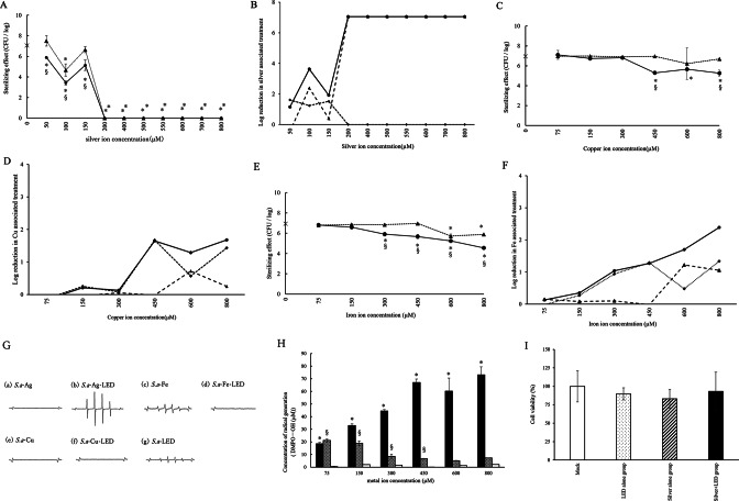
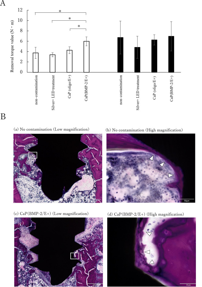
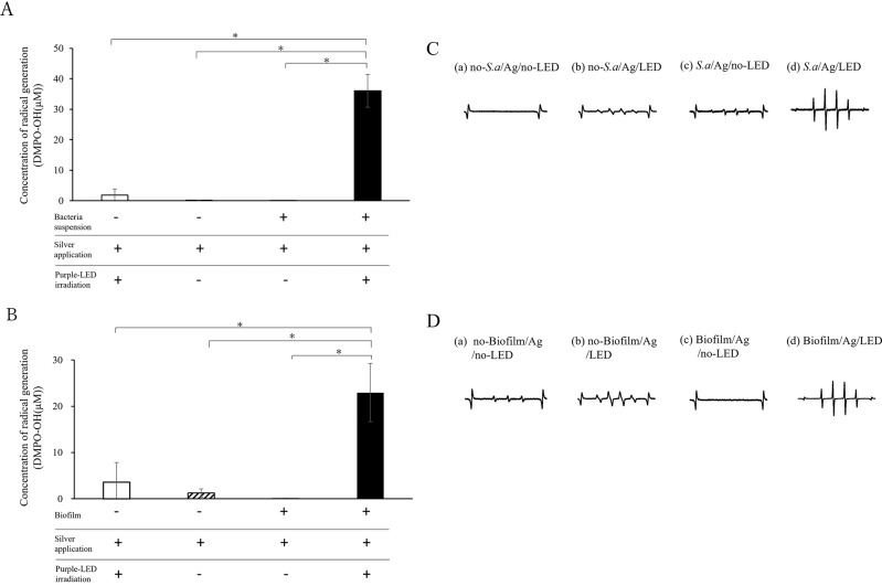
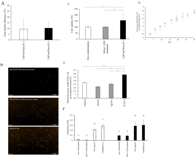
The aim of this study was to investigate the bactericidal effect and recovery of biocompatibility of contaminated titanium surfaces using a combination treatment involving silver, copper, or iron ion application along with 400 nm purple-LED light irradiation. Additionally, the study sought to develop a functional calcium phosphate (CaP) coating treatment on titanium surfaces following disinfection, to promote re-osseointegration. A purple-LED emitting light at 400 nm was utilized to irradiate Staphylococcus aureus suspensions and biofilms in the presence of various concentrations of silver, copper, and iron solutions for 1 min. The bactericidal effect and electron spin resonance (ESR) spectrum were subsequently evaluated. Additionally, the hydrophilicity of the titanium surface and cell viability of MC3T3-E1 cells after combination treatment with silver ion was evaluated. Furthermore, a titanium surface coating with CaP gene transfection carrier containing plasmid DNA was developed using an electric current. The activity of hard tissue formation was then evaluated both in vitro and in vivo post-treatment. The bactericidal effect of the combination treatment with silver ions was attributed to the generation of hydroxyl radicals, whereas the effects from iron and copper treatments were not radical-mediated. The silver treatment significantly restored the hydrophilicity and cell affinity of the titanium surface. Moreover, CaP coating applied via an electric current (30 µA for 5 min) enhanced hard tissue formation activity on the titanium surface in both in vitro and in vivo settings. The combination treatment utilizing silver ions and purple-LED irradiation significantly enhanced bactericidal effects by generating high levels of hydroxyl radicals. Additionally, coating the titanium surface with functionalized CaP promoted early osseointegration, suggesting a promising strategy for improving implant outcomes.
本研究的目的是探讨采用银、铜或铁离子应用与400纳米紫色发光二极管(LED)光照射相结合的联合处理方法,对受污染钛表面的杀菌效果及生物相容性的恢复情况。此外,该研究还试图在消毒后对钛表面进行功能性磷酸钙(CaP)涂层处理,以促进再骨整合。使用发出400纳米光的紫色LED在不同浓度的银、铜和铁溶液存在下照射金黄色葡萄球菌悬浮液和生物膜1分钟。随后评估杀菌效果和电子自旋共振(ESR)光谱。此外,还评估了银离子联合处理后钛表面的亲水性以及MC3T3-E1细胞的细胞活力。此外,利用电流开发了一种含有质粒DNA的CaP基因转染载体的钛表面涂层。然后在处理后的体外和体内评估硬组织形成活性。银离子联合处理的杀菌作用归因于羟基自由基的产生,而铁和铜处理的作用并非由自由基介导。银处理显著恢复了钛表面的亲水性和细胞亲和力。此外,通过电流(30微安,持续5分钟)施加的CaP涂层在体外和体内环境中均增强了钛表面的硬组织形成活性。银离子与紫色LED照射相结合的联合处理通过产生高水平的羟基自由基显著增强了杀菌效果。此外,用功能化的CaP对钛表面进行涂层促进了早期骨整合,这表明这是一种改善植入物效果的有前景的策略。