Parsons B W, Bedford M R, Wyatt C L
Department of Poultry Science, University of Arkansas at Fayetteville, AR 72701, USA.
AB Vista, Marlborough, United Kingdom.
Poult Sci. 2025 Apr 13;104(7):105161. doi: 10.1016/j.psj.2025.105161.
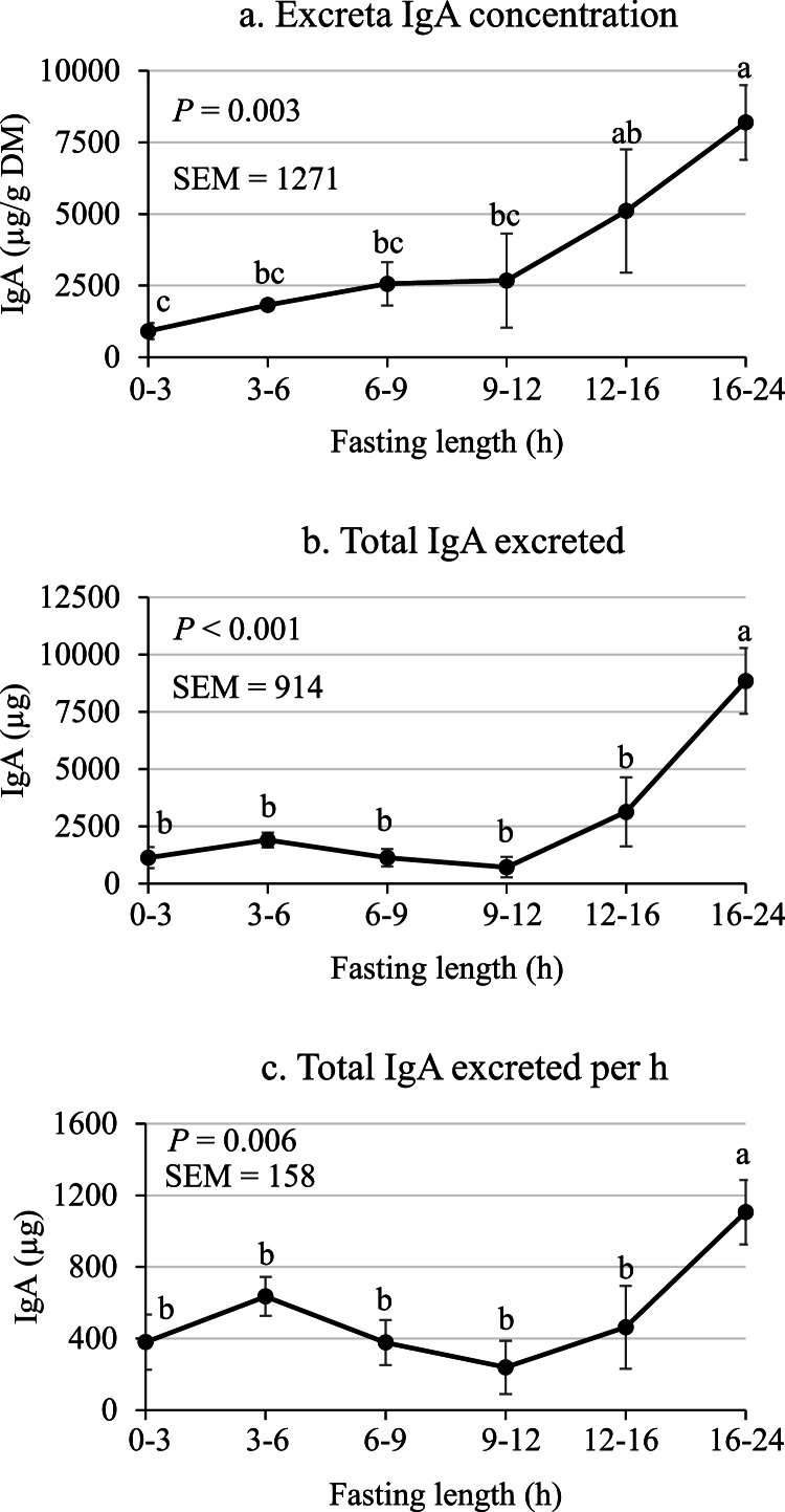
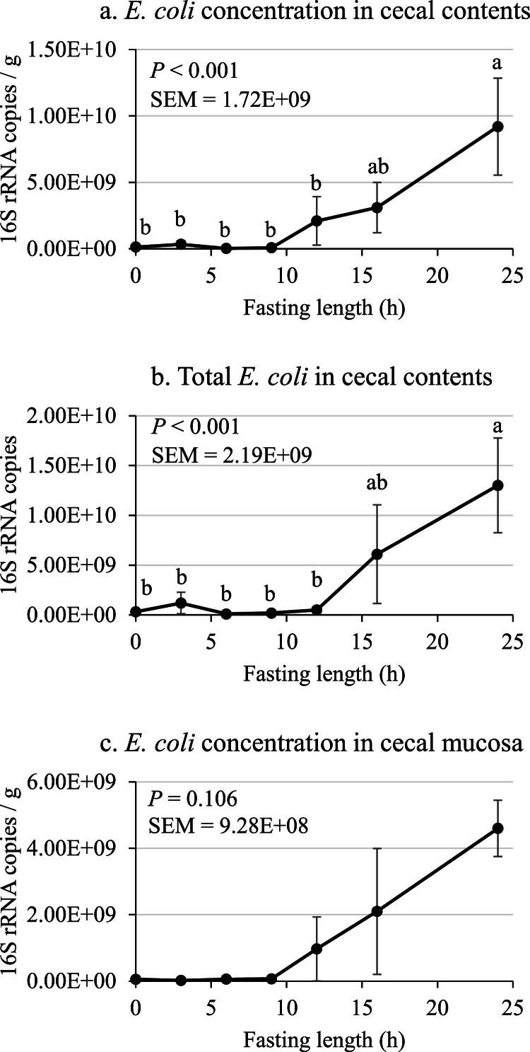
Two experiments were conducted to evaluate the effect of incremental fasting time on the gastrointestinal tract in chickens. Adult White Leghorn roosters with intact ceca were provided a nutrient-adequate corn-soybean meal diet ad libitum for 3 weeks. Prior to initiation of the experimental phase, ad libitum feed intake was recorded for 8 h and immediately after the fasting period commenced. In Experiment 1, roosters were fasted for either 0, 3, 6, 9, 12, 16, or 24 h. At each time point birds were euthanized and pH in the crop, gizzard, and ceca were recorded and cecal contents were collected to measure volatile fatty acids (VFA) and select cecal microbial groups. In Experiment 2, roosters were fasted for 0, 3, 6, 9, 12, 16, and 24 h and excreta were collected to determine secretory IgA (sIgA) excretion. In contrast to Experiment 1, roosters in Experiment 2 were not euthanized and thus sIgA excretion was measured within individual roosters across each time point. Experiments 1 and 2 contained 5 and 8 replicates per treatment, respectively. In Experiment 1, there was a linear increase (P < 0.05) in cecal pH as fasting length increased. Cecal VFA content was reduced (P < 0.05) by 9 to 12 h of fasting and branch-chain FA to VFA ratio increased (P < 0.05) by 6 h of fasting. There were few effects (P > 0.05) of fasting on the microbial groups in cecal contents and mucosa; however, Escherichia coli content was greater (P < 0.05) at 24 h of fasting compared with other time points. In Experiment 2, total sIgA excreted was greater (P < 0.05) at 24 h of fasting, being 1106 µg/h at 24 h compared with a mean of 419 µg/h for all other time points. In conclusion, fasting reduced cecal VFA concentrations and increased cecal pH, Escherichia coli, branched-chain FA to VFA ratio, and sIgA excretion, suggesting that fasting elicited negative effects on the gastrointestinal tract.
进行了两项实验以评估递增禁食时间对鸡胃肠道的影响。为成年白来航公鸡提供营养充足的玉米-豆粕日粮,自由采食3周。在实验阶段开始前,记录自由采食8小时的采食量以及禁食期开始后立即的采食量。在实验1中,公鸡分别禁食0、3、6、9、12、16或24小时。在每个时间点,对鸡实施安乐死,并记录嗉囊、砂囊和盲肠的pH值,收集盲肠内容物以测量挥发性脂肪酸(VFA)并选择盲肠微生物群。在实验2中,公鸡禁食0、3、6、9、12、16和24小时,并收集排泄物以测定分泌型IgA(sIgA)排泄量。与实验1不同,实验2中的公鸡未实施安乐死,因此在每个时间点的个体公鸡内测量sIgA排泄量。实验1和实验2中每个处理分别有5个和8个重复。在实验1中,随着禁食时间延长,盲肠pH值呈线性增加(P<0.05)。禁食9至12小时可降低盲肠VFA含量(P<0.05),禁食6小时可使支链脂肪酸与VFA的比例增加(P<0.05)。禁食对盲肠内容物和黏膜中的微生物群影响较小(P>0.05);然而,与其他时间点相比,禁食24小时时大肠杆菌含量更高(P<0.05)。在实验2中,禁食24小时时总sIgA排泄量更大(P<0.05),24小时时为1106μg/小时,而所有其他时间点的平均值为419μg/小时。总之,禁食降低了盲肠VFA浓度,增加了盲肠pH值、大肠杆菌、支链脂肪酸与VFA的比例以及sIgA排泄量,表明禁食对胃肠道产生了负面影响。