Wo Beibei, Liu Shuang, Liang Zihui, Li Xiaoming
Department of Otolaryngology Head and Neck Surgery, Hebei Medical University, Shijiazhuang, Hebei 050011, P.R. China.
Department of Pathology, The 980th Hospital of People's Liberation Army (PLA) Joint Logistics Support Force, Shijiazhuang, Hebei 050082, P.R. China.
Mol Med Rep. 2025 Jul;32(1). doi: 10.3892/mmr.2025.13548. Epub 2025 Apr 25.
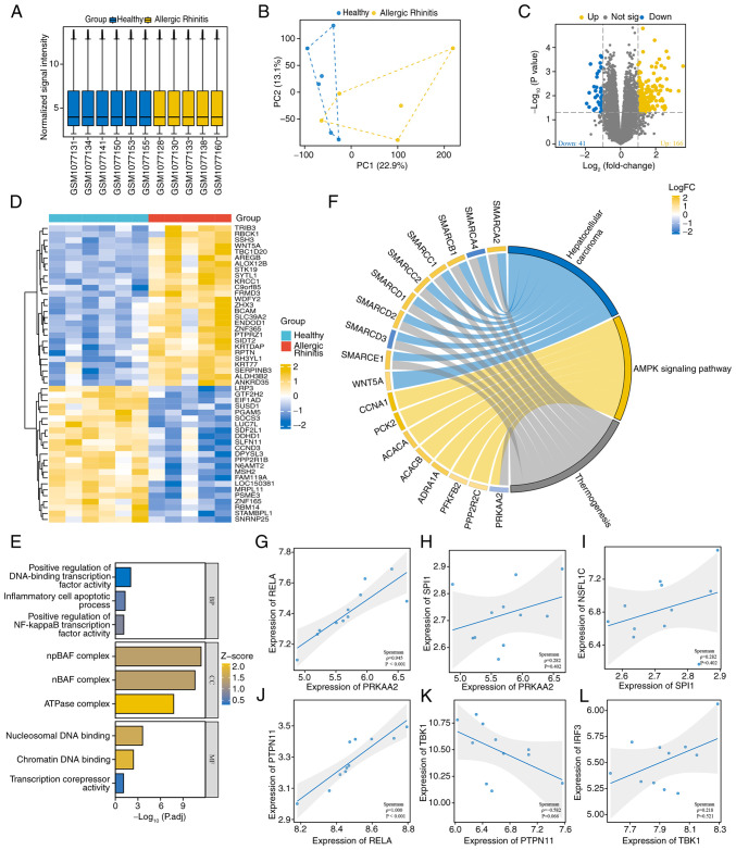
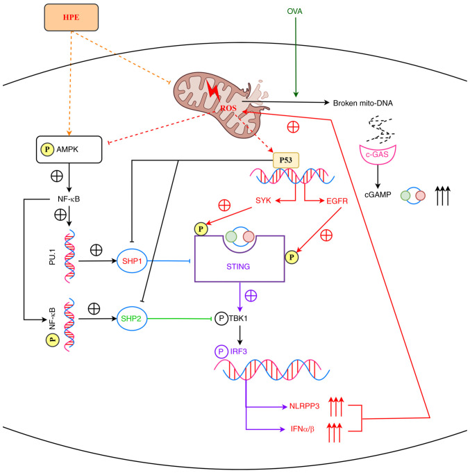
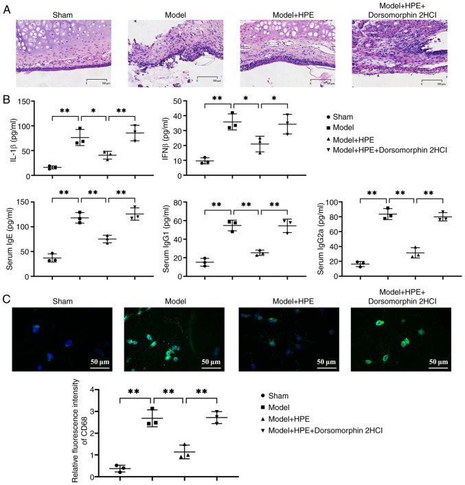
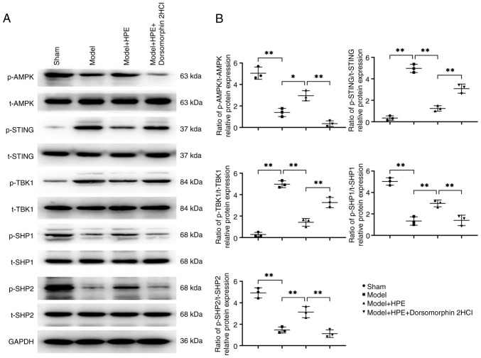
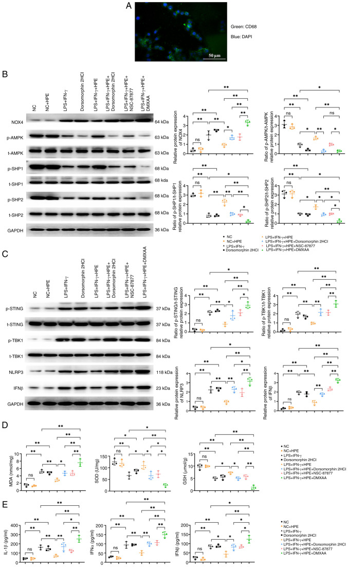
The present study aimed to investigate the regulatory effects and mechanisms of human placental extracts (HPE) on rats and cell models of ovalbumin (OVA)‑induced allergic rhinitis (AR). IFN‑y and LPS induced AR in vitro. A total of 32 male Sprague‑Dawley (SD) rats were randomly divided into the following four groups: Sham group, model group, model + HPE group and model + HPE + AMPK inhibitor group (n=8 rats/group). With the exception of the sham group, the remaining three groups were sensitized with OVA to establish an AR model, followed by various treatments. Hematoxylin and eosin staining was utilized to observe morphological changes in the nasal mucosa, ELISA was employed to measure serum levels of IL‑1β, interferon (IFN)β, immunoglobulin (Ig)E, IgG1 and IgG2a, and western blotting was conducted to assess protein expression across the groups. The sham group exhibited intact tissue structure with no notable pathological alterations. The model group demonstrated pronounced pathological features, including extensive infiltration of inflammatory cells, tissue shedding and edema. The model + HPE group revealed a gradual restoration of tissue architecture, characterized by reduced edema and inflammatory infiltration, whereas the model + HPE + AMPK inhibitor group again exhibited significant inflammatory cell infiltration and other pathological manifestations. Compared with the sham operation group, the levels of IL‑1β, IFNβ, IgE, IgG1 and IgG2a in the serum of the model group were elevated. The levels of IL‑1β, IFNβ, IgE, IgG1 and IgG2a in the model + HPE group were lower than those in the model group. In addition, the levels of IL‑1β, IFNβ, IgE, IgG1 and IgG2a in the model + HPE + AMPK inhibitor group were higher than those in the model + HPE group. Relative to the sham group, the expression levels of phosphorylated (p)‑AMPK/total (t)‑AMPK, p‑Src homology 2‑containing phosphatase (SHP)1/t‑SHP1 and p‑SHP2/t‑SHP2 were diminished, whereas the expression levels of p‑STING/t‑STING and p‑TBK1/t‑TBK1 were heightened in the model group. In comparison to the model group, the expression levels of p‑AMPK/t‑AMPK, p‑SHP1/t‑SHP1 and p‑SHP2/t‑SHP2 were enhanced, whereas the expression levels of p‑STING/t‑STING and p‑TBK1/t‑TBK1 were reduced in the model + HPE group. Conversely, when compared with the model + HPE group, the expression levels of p‑AMPK/t‑AMPK, p‑SHP1/t‑SHP1 and p‑SHP2/t‑SHP2 were decreased, whereas those of p‑STING/t‑STING and p‑TBK1/t‑TBK1 were increased in the model + HPE + AMPK inhibitor group. In conclusion, HPE may inhibit AR by modulating the AMPK/SHP1/SHP2/STING signaling pathway.
本研究旨在探讨人胎盘提取物(HPE)对卵清蛋白(OVA)诱导的大鼠变应性鼻炎(AR)及细胞模型的调节作用和机制。IFN-γ和LPS在体外诱导AR。将32只雄性Sprague-Dawley(SD)大鼠随机分为以下四组:假手术组、模型组、模型+HPE组和模型+HPE+AMPK抑制剂组(每组n = 8只大鼠)。除假手术组外,其余三组用OVA致敏以建立AR模型,然后进行各种处理。采用苏木精-伊红染色观察鼻黏膜的形态变化,采用酶联免疫吸附测定法(ELISA)检测血清白细胞介素-1β(IL-1β)、干扰素(IFN)β、免疫球蛋白(Ig)E、IgG1和IgG2a水平,并进行蛋白质免疫印迹法检测各组的蛋白表达。假手术组组织结构完整,无明显病理改变。模型组表现出明显的病理特征,包括炎性细胞广泛浸润、组织脱落和水肿。模型+HPE组显示组织结构逐渐恢复,其特征为水肿减轻和炎性浸润减少,而模型+HPE+AMPK抑制剂组再次出现明显的炎性细胞浸润和其他病理表现。与假手术组相比,模型组血清中IL-1β、IFNβ、IgE、IgG1和IgG2a水平升高。模型+HPE组中IL-1β、IFNβ、IgE、IgG1和IgG2a水平低于模型组。此外,模型+HPE+AMPK抑制剂组中IL-1β、IFNβ、IgE、IgG1和IgG2a水平高于模型+HPE组。相对于假手术组,模型组中磷酸化(p)-AMPK/总(t)-AMPK、p-含Src同源2结构域的磷酸酶(SHP)1/t-SHP1和p-SHP2/t-SHP2的表达水平降低,而p-干扰素基因刺激蛋白(STING)/t-STING和p-丝氨酸/苏氨酸蛋白激酶(TBK)1/t-TBK1的表达水平升高。与模型组相比,模型+HPE组中p-AMPK/t-AMPK、p-SHP1/t-SHP1和p-SHP2/t-SHP2的表达水平升高,而p-STING/t-STING和p-TBK1/t-TBK1的表达水平降低。相反,与模型+HPE组相比,模型+HPE+AMPK抑制剂组中p-AMPK/t-AMPK、p-SHP1/t-SHP1和p-SHP2/t-SHP2的表达水平降低,而p-STING/t-STING和p-TBK1/t-TBK1的表达水平升高。总之,HPE可能通过调节AMPK/SHP1/SHP2/STING信号通路抑制AR。