Dia Abdou Khadir, Kolnohuz Alona, Yolchuyeva Sevinj, Tonneau Marion, Lamaze Fabien, Orain Michele, Gagné Andréanne, Blais Florence, Coulombe François, Malo Julie, Belkaid Wiam, Elkrief Arielle, Williamson Drew, Routy Bertrand, Joubert Philippe, Laplante Mathieu, Bilodeau Steve, Manem Venkata Sk
Centre de Recherche du CHU de Québec - Université Laval, Québec, Canada.
Quebec Heart & Lung Institute Research Center, Québec, Canada.
J Transl Med. 2025 May 6;23(1):510. doi: 10.1186/s12967-025-06487-2.
Immune checkpoint inhibitors (ICIs) have revolutionized cancer treatment by significantly improving the efficacy of treatments and tolerability for patients with non-small cell lung cancer (NSCLC). However, even after meticulous selection based on molecular criteria, only 20-30% of the patients respond to ICIs. This highlights the urgent clinical need to develop more precise biomarkers to better identify individuals who will benefit from these expensive therapies.
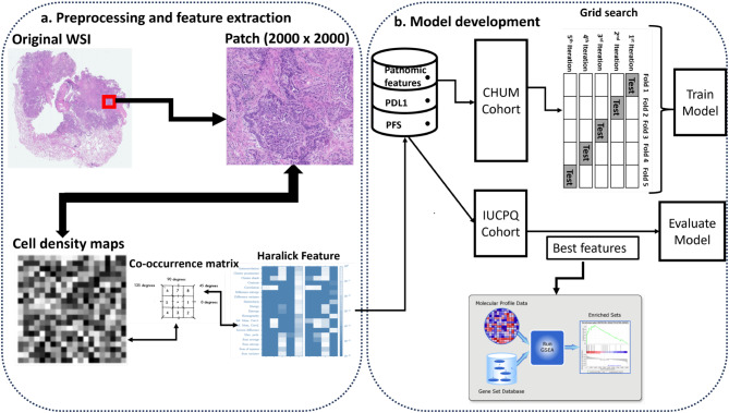
Data from NSCLC patients treated with immunotherapy were collected from two institutions. From the histological images of tumors, pathomics features were extracted. We employed six machine learning models and seven feature selection methods to predict expression of the programmed death-ligand 1 (PD-L1), a current biomarker used to select patients for immunotherapy, and progression-free survival (PFS). The association between pathomics features and biological pathways was explored to validate pathomics-based signatures. We performed gene set enrichment analysis to identify the pathways enriched with the predictive signatures.
Handcrafted histological features were extracted from the whole slide images (WSI). The Support Vector Machines model with the SurfStar feature selection method, offered the best results, with an area under the curve (AUC) of around 0.66 for both the training and validation sets to predict PD-L1. For the prediction of PFS, the most effective model was linear discriminant analysis using the Multi Surf feature selection method with an AUC of 0.71 for the training set and 0.62 for the validation set. We found immune pathways to be upregulated in the high PD-L1 and high PFS groups, confirming the utility of image analysis for predicting clinical endpoints in patients treated with immunotherapy.
Our models, based on the analysis of histological images, can serve as predictive biomarkers for PD-L1 and PFS. This approach, focused on histological images, enables the distinction of patients based on treatment response, thus providing clinicians with a valuable tool for patient management. With further validation on external cohorts, these models could enhance clinical decision-making through analysis of routine medical images.
免疫检查点抑制剂(ICI)通过显著提高非小细胞肺癌(NSCLC)患者的治疗效果和耐受性,彻底改变了癌症治疗方式。然而,即使根据分子标准进行了细致筛选,仍只有20%-30%的患者对ICI有反应。这凸显了迫切的临床需求,即开发更精确的生物标志物,以更好地识别将从这些昂贵疗法中获益的个体。
从两个机构收集接受免疫治疗的NSCLC患者的数据。从肿瘤的组织学图像中提取病理组学特征。我们采用六种机器学习模型和七种特征选择方法来预测程序性死亡配体1(PD-L1)的表达(一种目前用于选择免疫治疗患者的生物标志物)和无进展生存期(PFS)。探索病理组学特征与生物途径之间的关联,以验证基于病理组学的特征。我们进行基因集富集分析,以识别富含预测特征的途径。
从全切片图像(WSI)中提取手工制作的组织学特征。采用SurfStar特征选择方法的支持向量机模型给出了最佳结果,在训练集和验证集中预测PD-L1的曲线下面积(AUC)约为0.66。对于PFS的预测,最有效的模型是使用Multi Surf特征选择方法的线性判别分析,训练集的AUC为0.71,验证集的AUC为0.62。我们发现免疫途径在高PD-L1和高PFS组中上调,证实了图像分析在预测接受免疫治疗患者临床终点方面的实用性。
我们基于组织学图像分析的模型可作为PD-L1和PFS的预测生物标志物。这种专注于组织学图像的方法能够根据治疗反应区分患者,从而为临床医生提供了一个用于患者管理的有价值工具。通过在外部队列中进一步验证,这些模型可通过分析常规医学图像增强临床决策。