• 文献检索
  • 文档翻译
  • 深度研究
  • 学术资讯
  • Suppr Zotero 插件Zotero 插件
  • 邀请有礼
  • 套餐&价格
  • 历史记录
应用&插件
Suppr Zotero 插件Zotero 插件浏览器插件Mac 客户端Windows 客户端微信小程序
定价
高级版会员购买积分包购买API积分包
服务
文献检索文档翻译深度研究API 文档MCP 服务
关于我们
关于 Suppr公司介绍联系我们用户协议隐私条款
关注我们

Suppr 超能文献

核心技术专利:CN118964589B侵权必究
粤ICP备2023148730 号-1Suppr @ 2026

文献检索

告别复杂PubMed语法,用中文像聊天一样搜索,搜遍4000万医学文献。AI智能推荐,让科研检索更轻松。

立即免费搜索

文件翻译

保留排版,准确专业,支持PDF/Word/PPT等文件格式,支持 12+语言互译。

免费翻译文档

深度研究

AI帮你快速写综述,25分钟生成高质量综述,智能提取关键信息,辅助科研写作。

立即免费体验

使用三维姿势描记法评估手术前后腰骶部脊柱曲度。

Assessment of lumbosacral spinal curvatures before and after surgery using 3D posturography.

作者信息

Łosiński Karol, Bryndal Aleksandra, Grochulska Agnieszka, Nawos-Wysocki Wojciech, Glowinski Sebastian

机构信息

Institute of Health Sciences, Pomeranian University in Slupsk, Westerplatte 64, 76-200, Slupsk, Poland.

State Higher School of Vocational Education in Koszalin, Koszalin, Poland.

出版信息

Sci Rep. 2025 May 8;15(1):16126. doi: 10.1038/s41598-025-00510-8.

DOI:10.1038/s41598-025-00510-8
PMID:40341540
原文链接:https://pmc.ncbi.nlm.nih.gov/articles/PMC12062435/
Abstract

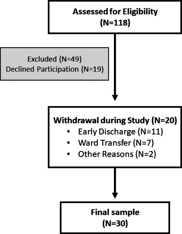
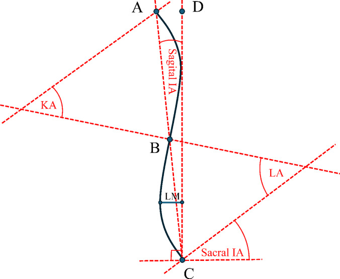
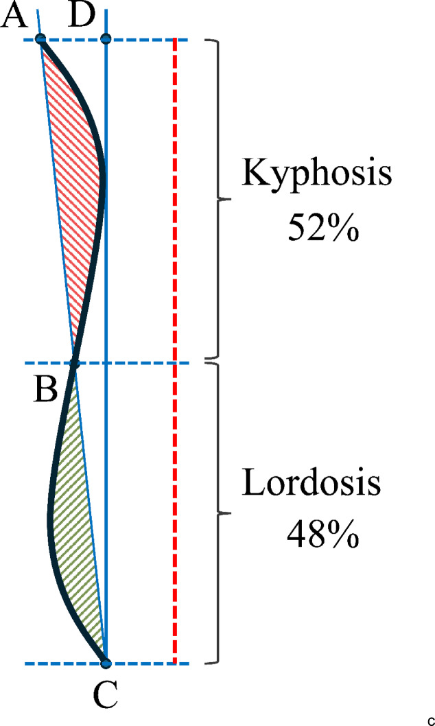
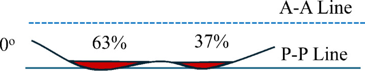
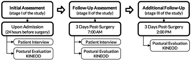
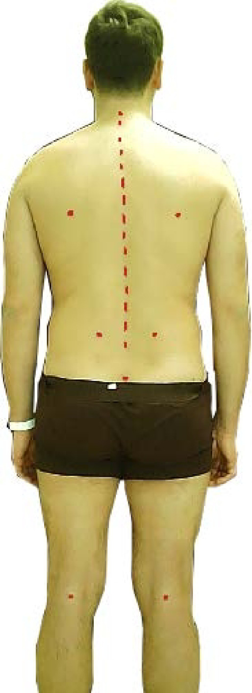
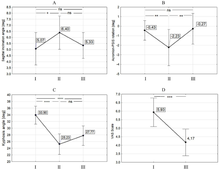
Open discectomy remains the standard procedure for decompressing neural structures in cases of intervertebral disc herniation. Early postoperative rehabilitation emphasizes protecting the surgical site while restoring functional independence in daily activities. In the absence of possibilities for performing any spinal control tests within the first few days post-surgery, the dynamism of curvature changes may be the sole criterion for evaluating rehabilitation progress. This study aims to evaluate the feasibility of utilizing the non-invasive KINEOD device to measure the dynamics of lumbosacral spinal alignment parameters in the sagittal plane before and immediately after surgical intervention. The study involved 30 patients (14 women [46.67%] and 16 men [53.33%]) from the Neurosurgical Department of the Wojewódzki Szpital Specjalistyczny in Słupsk, Poland. Data were collected using a custom questionnaire and KINEOD 3D posturography to assess body posture. The following parameters were analyzed: Sagittal Inclination Angle (Sagittal IA), Kyphosis Angle (KA), Lordosis Angle (LA), Sacral Inclination Angle (Sacral IA), Lordosis Measure (LM), Inflection Point (IP), and the Angle of Acromion-Posterior Superior Iliac Spine Rotation (A-PSIS). Measurements were conducted at three stages: Stage I - one day before surgery (2:00 PM); Stage II - on the third postoperative day (7:00 AM); and Stage III - on the third postoperative day (2:00 PM). Measurements utilizing the KINEOD device revealed statistically significant changes between Stage I and Stage II for the following parameters: Sagittal IA, KA, LA, Sacral IA, LM, IP, and A-PSIS. Significant changes were also noted between Stage I and Stage III for KA, LA, Sacral IA, LM, A-PSIS, and VAS. The study highlights that the reliability of KINEOD 3D assessment diminishes when relying solely on parameters such as Sacral IA and IP for postoperative lordosis evaluation. For rapid, non-invasive assessment of the lumbosacral spine post-surgery, Sacral IA may serve as a more accurate indicator of dynamic changes in the lower lumbar region. Postoperative alterations in all sagittal plane angles are influenced by both surgical intervention and, to a lesser extent, the diurnal adaptation rhythm.

摘要

对于椎间盘突出症患者,开放式椎间盘切除术仍然是减压神经结构的标准手术。术后早期康复强调在保护手术部位的同时,恢复日常活动中的功能独立性。在术后头几天无法进行任何脊柱控制测试的情况下,脊柱曲度变化的动态情况可能是评估康复进展的唯一标准。本研究旨在评估使用非侵入性KINEOD设备测量手术干预前后矢状面腰骶部脊柱排列参数动态变化的可行性。该研究纳入了波兰斯武普斯克 Wojewódzki Szpital Specjalistyczny神经外科的30例患者(14名女性[46.67%]和16名男性[53.33%])。通过定制问卷和KINEOD 3D姿势描记法收集数据以评估身体姿势。分析了以下参数:矢状面倾斜角(Sagittal IA)、后凸角(KA)、前凸角(LA)、骶骨倾斜角(Sacral IA)、前凸测量值(LM)、拐点(IP)以及肩峰-后上棘旋转角(A-PSIS)。测量在三个阶段进行:第一阶段——手术前一天下午2点;第二阶段——术后第三天上午7点;第三阶段——术后第三天下午2点。使用KINEOD设备进行的测量显示,在第一阶段和第二阶段之间,以下参数存在统计学上的显著变化:矢状面IA、KA、LA、骶骨IA、LM、IP和A-PSIS。在第一阶段和第三阶段之间,KA、LA、骶骨IA、LM、A-PSIS和视觉模拟评分(VAS)也有显著变化。该研究强调,仅依靠骶骨IA和IP等参数进行术后前凸评估时,KINEOD 3D评估的可靠性会降低。对于术后腰骶部脊柱的快速、非侵入性评估,骶骨IA可能是下腰椎区域动态变化更准确的指标。矢状面所有角度的术后改变均受手术干预影响,且在较小程度上受昼夜适应节律影响。

https://cdn.ncbi.nlm.nih.gov/pmc/blobs/5c2f/12062435/6392accbf06a/41598_2025_510_Fig10_HTML.jpg
https://cdn.ncbi.nlm.nih.gov/pmc/blobs/5c2f/12062435/190d41fa65c1/41598_2025_510_Fig1_HTML.jpg
https://cdn.ncbi.nlm.nih.gov/pmc/blobs/5c2f/12062435/1e097ec3e411/41598_2025_510_Fig2_HTML.jpg
https://cdn.ncbi.nlm.nih.gov/pmc/blobs/5c2f/12062435/db61536a72f8/41598_2025_510_Fig3_HTML.jpg
https://cdn.ncbi.nlm.nih.gov/pmc/blobs/5c2f/12062435/c021dfa1c712/41598_2025_510_Fig4_HTML.jpg
https://cdn.ncbi.nlm.nih.gov/pmc/blobs/5c2f/12062435/1b9995e0e1eb/41598_2025_510_Fig5_HTML.jpg
https://cdn.ncbi.nlm.nih.gov/pmc/blobs/5c2f/12062435/ccd12bd1da85/41598_2025_510_Fig6_HTML.jpg
https://cdn.ncbi.nlm.nih.gov/pmc/blobs/5c2f/12062435/5f293f7ac0fa/41598_2025_510_Fig7_HTML.jpg
https://cdn.ncbi.nlm.nih.gov/pmc/blobs/5c2f/12062435/059dca30fa12/41598_2025_510_Fig8_HTML.jpg
https://cdn.ncbi.nlm.nih.gov/pmc/blobs/5c2f/12062435/deee82605832/41598_2025_510_Fig9_HTML.jpg
https://cdn.ncbi.nlm.nih.gov/pmc/blobs/5c2f/12062435/6392accbf06a/41598_2025_510_Fig10_HTML.jpg

相似文献

1
Assessment of lumbosacral spinal curvatures before and after surgery using 3D posturography.使用三维姿势描记法评估手术前后腰骶部脊柱曲度。
Sci Rep. 2025 May 8;15(1):16126. doi: 10.1038/s41598-025-00510-8.
2
Progressive restoration of spinal sagittal balance after surgical correction of lumbosacral spondylolisthesis before skeletal maturity.骨骼成熟前腰骶部椎体滑脱手术矫正后脊柱矢状面平衡的渐进性恢复
J Neurosurg Spine. 2015 Mar;22(3):294-300. doi: 10.3171/2014.9.SPINE1412. Epub 2015 Jan 9.
3
Influence of cage geometry on sagittal alignment in instrumented posterior lumbar interbody fusion.椎间融合器几何形状对后路腰椎椎间融合内固定矢状面排列的影响。
Spine (Phila Pa 1976). 2003 Aug 1;28(15):1693-9. doi: 10.1097/01.BRS.0000083167.78853.D5.
4
Measurements of lumbopelvic lordosis using the pelvic radius technique as it correlates with sagittal spinal balance and sacral translation.采用骨盆半径技术测量腰骶部前凸,因为它与矢状面脊柱平衡和骶骨移位相关。
Spine J. 2002 Nov-Dec;2(6):421-9. doi: 10.1016/s1529-9430(02)00426-6.
5
Spinal sagittal imbalance in patients with lumbar disc herniation: its spinopelvic characteristics, strength changes of the spinal musculature and natural history after lumbar discectomy.腰椎间盘突出症患者的脊柱矢状面失衡:其脊柱骨盆特征、脊柱肌肉力量变化及腰椎间盘切除术后的自然病程。
BMC Musculoskelet Disord. 2016 Jul 22;17:305. doi: 10.1186/s12891-016-1164-y.
6
Interpretation of Spinal Radiographic Parameters in Patients With Transitional Lumbosacral Vertebrae.过渡性腰骶椎患者脊柱影像学参数的解读
Spine Deform. 2018 Sep-Oct;6(5):587-592. doi: 10.1016/j.jspd.2018.01.004.
7
Characterization of Sagittal Spine Alignment With Reference to the Gravity Line and Vertebral Slopes: An Analysis of Different Roussouly Curves.基于重力线和椎体斜率评估矢状位脊柱排列:不同 Roussouly 曲线分析。
Spine (Phila Pa 1976). 2020 May 1;45(9):E481-E488. doi: 10.1097/BRS.0000000000003379.
8
Influence of lumbar lordosis restoration on thoracic curve and sagittal position in lumbar degenerative kyphosis patients.腰椎前凸恢复对腰椎退行性后凸患者胸椎曲度及矢状位的影响。
Spine (Phila Pa 1976). 2009 Feb 1;34(3):280-4. doi: 10.1097/BRS.0b013e318191e792.
9
Sagittal spinal alignment in patients with lumbar disc herniation.腰椎间盘突出症患者的矢状位脊柱排列。
Eur Spine J. 2010 Mar;19(3):435-8. doi: 10.1007/s00586-009-1240-1. Epub 2009 Dec 20.
10
Changes in the lumbosacral angle, sacral inclination and the curvature of the lumbar spine during aging.衰老过程中腰骶角、骶骨倾斜度及腰椎曲度的变化。
Acta Anat (Basel). 1992;145(4):373-7. doi: 10.1159/000147392.

引用本文的文献

1
Traumatic Bilateral Lumbosacral Jumped Facet Without Fracture in Childhood: Case Report and Systematic Review.儿童期创伤性双侧腰骶关节突关节脱位无骨折:病例报告及系统评价
J Clin Med. 2025 Sep 3;14(17):6228. doi: 10.3390/jcm14176228.

本文引用的文献

1
Experimental ex vivo characterization of the biomechanical effects of laminectomy and posterior fixation of the lumbo-sacral spine.腰椎-骶椎椎板切除术及后路固定生物力学效应的体外实验表征
Sci Rep. 2024 Dec 3;14(1):30001. doi: 10.1038/s41598-024-80741-3.
2
Risk factors and treatment strategies for adjacent segment disease following spinal fusion (Review).脊柱融合术后邻近节段病的危险因素和治疗策略(综述)。
Mol Med Rep. 2025 Feb;31(2). doi: 10.3892/mmr.2024.13398. Epub 2024 Nov 22.
3
Outcomes and Spinopelvic Changes After Anterior Lumbar Interbody Fusion With a Novel Interbody Fusion Device: A Retrospective Study.
使用新型椎间融合器进行腰椎前路椎间融合术后的疗效及脊柱骨盆变化:一项回顾性研究
Int J Spine Surg. 2024 Sep 12;18(4):389-399. doi: 10.14444/8621.
4
Positioning the surgical patient - roles, responsibilities and challenges. A qualitative study.手术患者的体位摆放——角色、职责与挑战。一项定性研究。
J Adv Nurs. 2025 Feb;81(2):968-977. doi: 10.1111/jan.16278. Epub 2024 Jun 18.
5
Change in Lumbar Lordosis After Decompressive Surgery in Lumbar Spinal Stenosis Patients and Associations With Patient-Related Outcomes Two Years After Surgery: Radiologic and Clinical Results From the NORDSTEN Spinal Stenosis Trial.腰椎管狭窄症患者减压手术后腰椎前凸的变化及其与术后两年患者相关结局的关联:NORDSTEN腰椎管狭窄症试验的影像学和临床结果
Spine (Phila Pa 1976). 2025 Jan 1;50(1):40-45. doi: 10.1097/BRS.0000000000005037. Epub 2024 May 13.
6
Use of a Triaxial Accelerometer to Measure Changes in Gait Sway and Related Motor Function after Corrective Spinal Fusion Surgery for Adult Spinal Deformity.使用三轴加速度计测量成人脊柱畸形矫正性脊柱融合手术后步态摆动及相关运动功能的变化。
J Clin Med. 2024 Mar 26;13(7):1923. doi: 10.3390/jcm13071923.
7
Rehabilitation after cervical and lumbar spine surgery.颈椎和腰椎手术后的康复
EFORT Open Rev. 2023 Aug 1;8(8):626-638. doi: 10.1530/EOR-23-0015.
8
Intervertebral disc degeneration-Current therapeutic options and challenges.椎间盘退变——当前的治疗选择和挑战。
Front Public Health. 2023 Jul 6;11:1156749. doi: 10.3389/fpubh.2023.1156749. eCollection 2023.
9
Quantitative Assessment of Intervertebral Disc Composition by MRI: Sensitivity to Diurnal Variation.基于 MRI 的椎间盘成分定量评估:对昼夜变化的敏感性。
Tomography. 2023 May 16;9(3):1029-1040. doi: 10.3390/tomography9030084.
10
Increased cage angle effects on radiographic outcomes after stand-alone anterior lumbar interbody fusion.单纯前路腰椎间融合术后 cage 角度增加对影像学结果的影响。
J Neurosurg Spine. 2023 May 5;39(2):254-262. doi: 10.3171/2023.4.SPINE221283. Print 2023 Aug 1.