• 文献检索
  • 文档翻译
  • 深度研究
  • 学术资讯
  • Suppr Zotero 插件Zotero 插件
  • 邀请有礼
  • 套餐&价格
  • 历史记录
应用&插件
Suppr Zotero 插件Zotero 插件浏览器插件Mac 客户端Windows 客户端微信小程序
定价
高级版会员购买积分包购买API积分包
服务
文献检索文档翻译深度研究API 文档MCP 服务
关于我们
关于 Suppr公司介绍联系我们用户协议隐私条款
关注我们

Suppr 超能文献

核心技术专利:CN118964589B侵权必究
粤ICP备2023148730 号-1Suppr @ 2026

文献检索

告别复杂PubMed语法,用中文像聊天一样搜索,搜遍4000万医学文献。AI智能推荐,让科研检索更轻松。

立即免费搜索

文件翻译

保留排版,准确专业,支持PDF/Word/PPT等文件格式,支持 12+语言互译。

免费翻译文档

深度研究

AI帮你快速写综述,25分钟生成高质量综述,智能提取关键信息,辅助科研写作。

立即免费体验

从功能到结构:肌原纤维生成如何影响横向-轴向管状系统的发育及其特性。

From function to structure: how myofibrillogenesis influences the transverse-axial tubular system development and its peculiarities.

作者信息

Sevcikova Tomaskova Zuzana, Mackova Katarina

机构信息

Department of Biophysics and Electrophysiology, Institute of Molecular Physiology and Genetics, Centre of Biosciences, Slovak Academy of Sciences, Bratislava, Slovakia.

出版信息

Front Physiol. 2025 Apr 25;16:1576133. doi: 10.3389/fphys.2025.1576133. eCollection 2025.

DOI:10.3389/fphys.2025.1576133
PMID:40352140
原文链接:https://pmc.ncbi.nlm.nih.gov/articles/PMC12062141/
Abstract

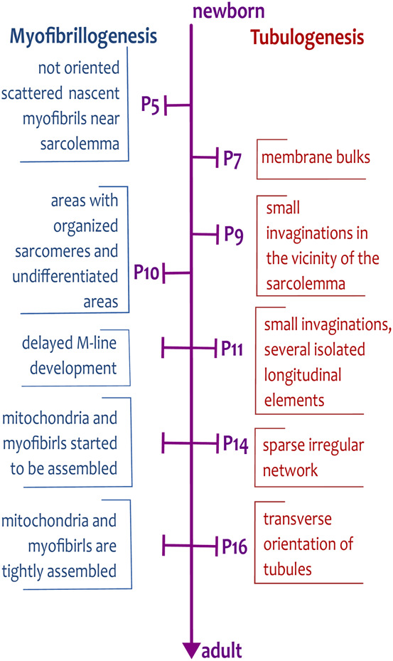
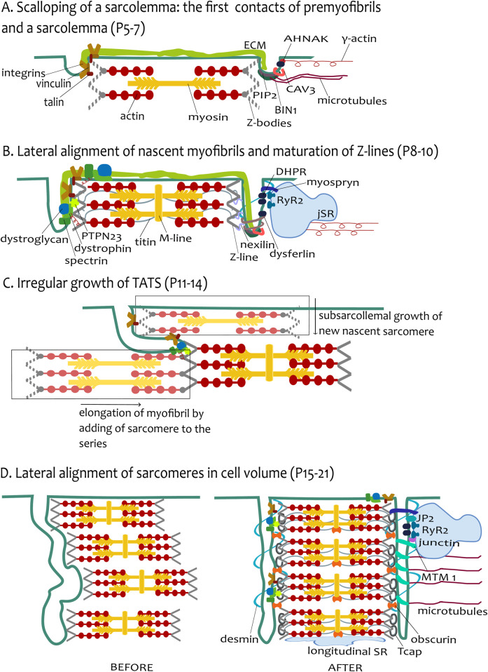
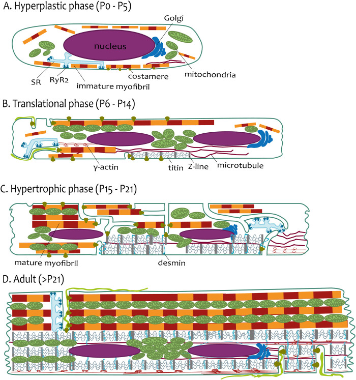
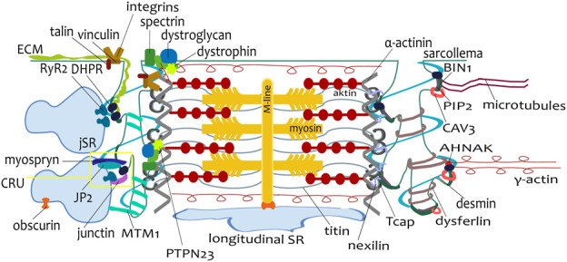
The transverse-axial tubular system (TATS) is the extension of sarcolemma growing to the cell interior, providing sufficient calcium signaling to induce calcium release from sarcoplasmic reticulum cisternae and stimulate the contraction of neighboring myofibrils. Interestingly, the development of TATS is delayed and matures during the period. It starts with small invaginations near the sarcolemma, proceeding to grow an irregular network that is later assembled into the notably transversally oriented tubular network. Accumulating evidence supports the idea that the development of TATS is linked to cell dimensions, calcium signaling, and increasing myofibrillar content orchestrated by electromechanical stimulation. However, the overall mechanism has not yet been described. The topic of this review is the development of TATS with an emphasis on the irregular phase of tubule growth. The traditional models of BIN1-related tubulation are also discussed. We summarized the recently described protein interactions during TATS development, mainly mediated by costameric and sarcomeric proteins, supporting the idea of the coupling sites between TATS and the myofibrils. We hypothesize that the formation and final organization of the tubular system is driven by the simultaneous development of the contractile apparatus under cycling electromechanical stimulus.

摘要

横-轴管状系统(TATS)是肌膜向细胞内部生长的延伸部分,可提供足够的钙信号,以诱导肌浆网池释放钙,并刺激相邻肌原纤维的收缩。有趣的是,TATS的发育在此期间延迟并成熟。它始于肌膜附近的小凹陷,进而发展成不规则网络,随后组装成明显横向排列的管状网络。越来越多的证据支持这样一种观点,即TATS的发育与细胞大小、钙信号以及由机电刺激精心安排的肌原纤维含量增加有关。然而,其整体机制尚未得到描述。本综述的主题是TATS的发育,重点是小管生长的不规则阶段。还讨论了与BIN1相关的微管形成的传统模型。我们总结了最近描述的TATS发育过程中的蛋白质相互作用,主要由肌附着蛋白和肌节蛋白介导,支持TATS与肌原纤维之间耦合位点的观点。我们假设管状系统的形成和最终组织是由循环机电刺激下收缩装置的同步发育驱动的。

https://cdn.ncbi.nlm.nih.gov/pmc/blobs/b6c5/12062141/e61cb4865684/fphys-16-1576133-g004.jpg
https://cdn.ncbi.nlm.nih.gov/pmc/blobs/b6c5/12062141/990c5ecbb766/fphys-16-1576133-g001.jpg
https://cdn.ncbi.nlm.nih.gov/pmc/blobs/b6c5/12062141/3747e5f5465c/fphys-16-1576133-g002.jpg
https://cdn.ncbi.nlm.nih.gov/pmc/blobs/b6c5/12062141/50fe9452b3e9/fphys-16-1576133-g003.jpg
https://cdn.ncbi.nlm.nih.gov/pmc/blobs/b6c5/12062141/e61cb4865684/fphys-16-1576133-g004.jpg

相似文献

1
From function to structure: how myofibrillogenesis influences the transverse-axial tubular system development and its peculiarities.从功能到结构:肌原纤维生成如何影响横向-轴向管状系统的发育及其特性。
Front Physiol. 2025 Apr 25;16:1576133. doi: 10.3389/fphys.2025.1576133. eCollection 2025.
2
Different effects of cardiomyocyte contractile activity on transverse and axial tubular system luminal content dynamics.心肌细胞收缩活动对横向和轴向管状系统管腔内容物动力学的不同影响。
J Mol Cell Cardiol. 2024 Dec;197:125-135. doi: 10.1016/j.yjmcc.2024.10.014. Epub 2024 Nov 2.
3
Mechanisms of spontaneous Ca release-mediated arrhythmia in a novel 3D human atrial myocyte model: I. Transverse-axial tubule variation.新型三维人心房肌细胞模型中自发性 Ca 释放介导的心律失常机制:I. 横-纵管变异性。
J Physiol. 2023 Jul;601(13):2655-2683. doi: 10.1113/JP283363. Epub 2022 Oct 27.
4
Role of the transverse-axial tubule system in generating calcium sparks and calcium transients in rat atrial myocytes.横-轴管系统在大鼠心房肌细胞中产生钙火花和钙瞬变的作用。
J Physiol. 2003 Mar 1;547(Pt 2):441-51. doi: 10.1113/jphysiol.2002.034355. Epub 2003 Jan 31.
5
Quantitative assessment of passive electrical properties of the cardiac T-tubular system by FRAP microscopy.荧光相关光漂白显微镜定量评估心脏 T 管系统的被动电特性。
Proc Natl Acad Sci U S A. 2017 May 30;114(22):5737-5742. doi: 10.1073/pnas.1702188114. Epub 2017 May 15.
6
Modelling the cardiac transverse-axial tubular system.构建心脏横向轴管系统模型。
Prog Biophys Mol Biol. 2008 Jan-Apr;96(1-3):226-43. doi: 10.1016/j.pbiomolbio.2007.07.021. Epub 2007 Aug 11.
7
The transverse-axial tubular system of cardiomyocytes.心肌细胞的横管-纵管系统。
Cell Mol Life Sci. 2013 Dec;70(24):4695-710. doi: 10.1007/s00018-013-1410-5. Epub 2013 Jul 12.
8
Heterogeneity of transverse-axial tubule system in mouse atria: Remodeling in atrial-specific Na-Ca exchanger knockout mice.小鼠心房横向轴突小管系统的异质性:心房特异性钠钙交换体基因敲除小鼠中的重塑。
J Mol Cell Cardiol. 2017 Jul;108:50-60. doi: 10.1016/j.yjmcc.2017.05.008. Epub 2017 May 19.
9
Mechanisms of spontaneous Ca release-mediated arrhythmia in a novel 3D human atrial myocyte model: II. Ca -handling protein variation.新型三维人心房肌细胞模型中介导自发性钙释放的心律失常机制:Ⅱ. 钙处理蛋白的变化。
J Physiol. 2023 Jul;601(13):2685-2710. doi: 10.1113/JP283602. Epub 2022 Sep 29.
10
Development of the excitation-contraction coupling apparatus in skeletal muscle: peripheral and internal calcium release units are formed sequentially.骨骼肌兴奋-收缩偶联装置的发育:外周和内部钙释放单元依次形成。
J Muscle Res Cell Motil. 1994 Apr;15(2):102-18. doi: 10.1007/BF00130422.

本文引用的文献

1
Restoring Atrial T-Tubules Augments Systolic Ca Upon Recovery From Heart Failure.恢复心房 T 小管可增强心力衰竭恢复后的收缩期 Ca。
Circ Res. 2024 Sep 13;135(7):739-754. doi: 10.1161/CIRCRESAHA.124.324601. Epub 2024 Aug 14.
2
Matriglycan maintains t-tubule structural integrity in cardiac muscle.基质糖蛋白维持心肌中的 T 小管结构完整性。
Proc Natl Acad Sci U S A. 2024 May 28;121(22):e2402890121. doi: 10.1073/pnas.2402890121. Epub 2024 May 21.
3
Human induced pluripotent stem cell-derived closed-loop cardiac tissue for drug assessment.
用于药物评估的人诱导多能干细胞来源的闭环心脏组织。
iScience. 2024 Jan 23;27(2):108992. doi: 10.1016/j.isci.2024.108992. eCollection 2024 Feb 16.
4
Ptpn23 Controls Cardiac T-Tubule Patterning by Promoting the Assembly of Dystrophin-Glycoprotein Complex.Ptpn23通过促进肌营养不良蛋白-糖蛋白复合物的组装来控制心脏T小管的形态。
Circulation. 2024 Apr 23;149(17):1375-1390. doi: 10.1161/CIRCULATIONAHA.123.065767. Epub 2024 Jan 12.
5
BIN1, Myotubularin, and Dynamin-2 Coordinate T-Tubule Growth in Cardiomyocytes.BIN1、Myotubularin 和 Dynamin-2 协调心肌细胞中的 T 小管生长。
Circ Res. 2023 May 26;132(11):e188-e205. doi: 10.1161/CIRCRESAHA.122.321732. Epub 2023 May 4.
6
Caveolae and Bin1 form ring-shaped platforms for T-tubule initiation.陷窝小体和 Bin1 形成环形平台,用于 T 管起始。
Elife. 2023 Apr 21;12:e84139. doi: 10.7554/eLife.84139.
7
Engineering the maturation of stem cell-derived cardiomyocytes.调控干细胞来源心肌细胞的成熟
Front Bioeng Biotechnol. 2023 Mar 22;11:1155052. doi: 10.3389/fbioe.2023.1155052. eCollection 2023.
8
Membrane remodelling triggers maturation of excitation-contraction coupling in 3D-shaped human-induced pluripotent stem cell-derived cardiomyocytes.膜重塑触发三维人诱导多能干细胞衍生心肌细胞兴奋-收缩耦联的成熟。
Basic Res Cardiol. 2023 Mar 29;118(1):13. doi: 10.1007/s00395-023-00984-5.
9
Generation and maturation of human iPSC-derived 3D organotypic cardiac microtissues in long-term culture.人诱导多能干细胞衍生的 3D 器官型心脏微组织的长期培养中的生成和成熟。
Sci Rep. 2022 Oct 18;12(1):17409. doi: 10.1038/s41598-022-22225-w.
10
Serial block face scanning electron microscopy reveals region-dependent remodelling of transverse tubules post-myocardial infarction.序列块面扫描电子显微镜显示心肌梗死后横管的区域依赖性重塑。
Philos Trans R Soc Lond B Biol Sci. 2022 Nov 21;377(1864):20210331. doi: 10.1098/rstb.2021.0331. Epub 2022 Oct 3.