Matsuda Shinya, Saito Chieko, Nomura Mami, Kawahara Hitomi, Mizushima Noboru, Nakano Kentaro
Degree Programs in Biology, Graduate School of Science and Technology, University of Tsukuba, Tsukuba, Ibaraki Prefecture, Japan.
College of Biological Sciences, School of Life and Environmental Sciences, University of Tsukuba, Tsukuba, Ibaraki Prefecture, Japan.
mBio. 2025 Jun 11;16(6):e0078325. doi: 10.1128/mbio.00783-25. Epub 2025 May 15.
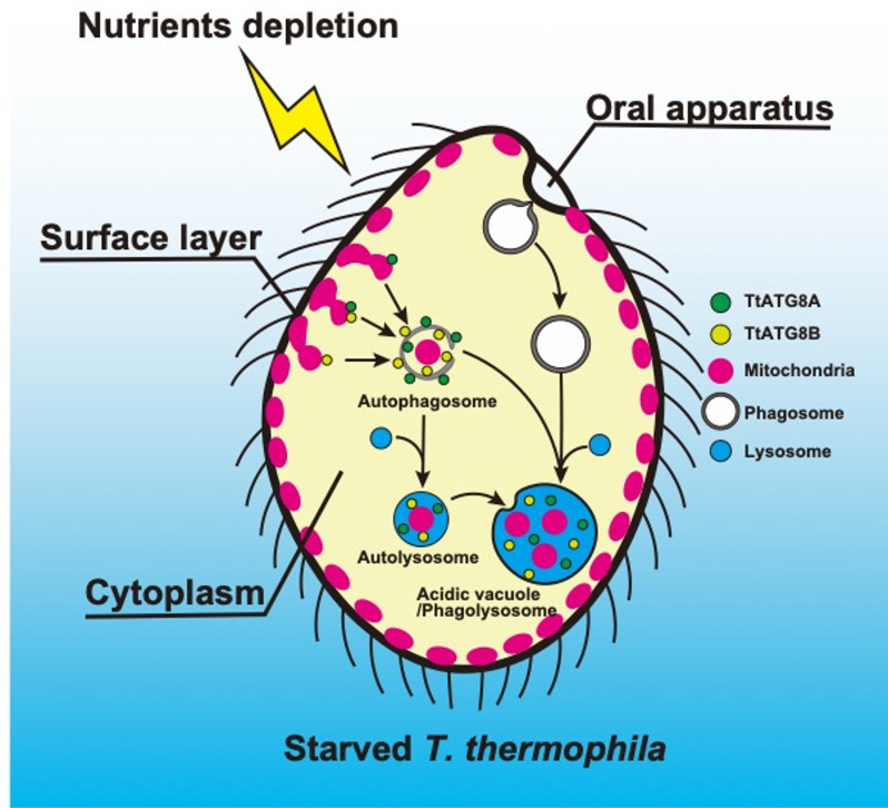
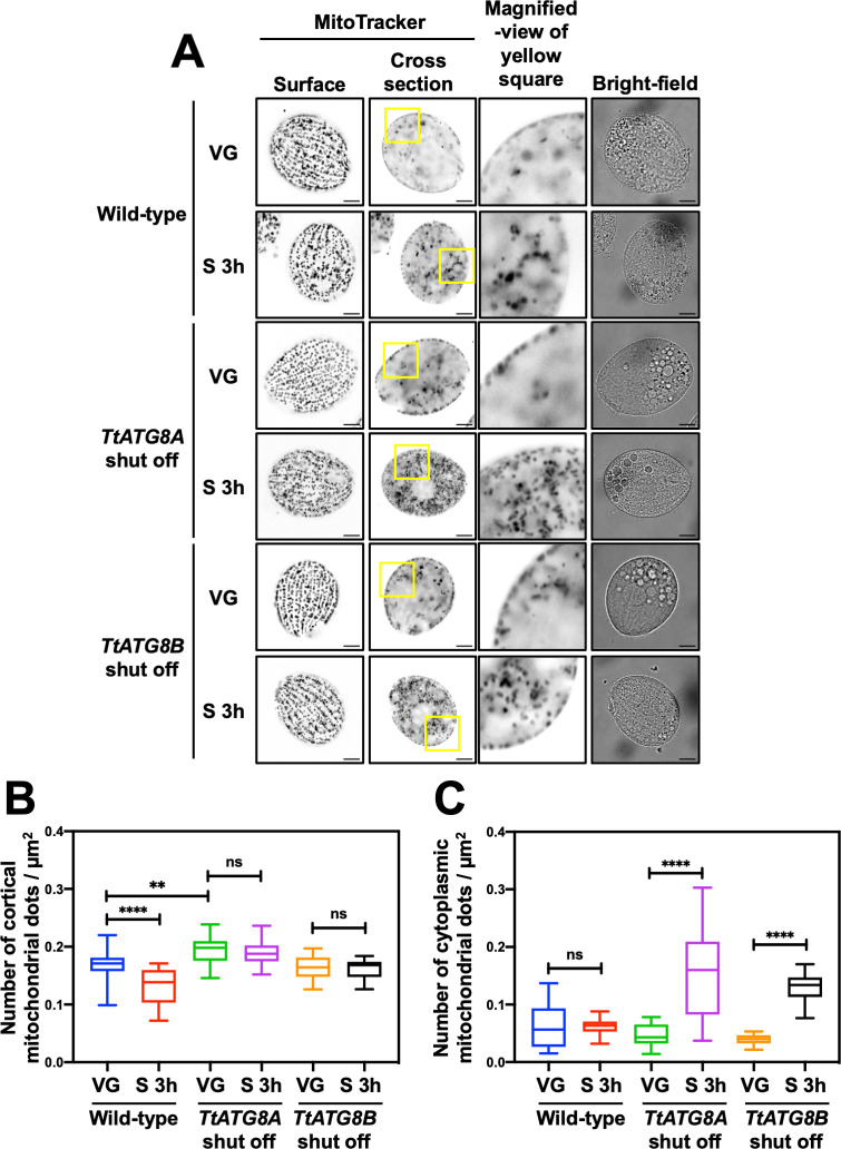
The majority of heterotrophic unicellular eukaryotes have evolved mechanisms to survive periods of starvation, allowing them to endure until conditions are favorable for regrowth. The ciliate exhibits active swimming behavior in water, preying on microorganisms and growing exponentially at a rate of 0.5-0.75 h⁻¹ under optimal conditions. In this organism, numerous mitochondria localize to the cell cortex along the ciliary rows, likely ensuring an efficient ATP supply necessary for vigorous cell movement. Although mitochondrial reduction occurs immediately under starvation, the underlying mechanism remains unknown. Here, we demonstrated that autophagy is responsible for mitochondrial reduction in . Among the five ATG8 homologs, TtATG8A and TtATG8B formed granule- and cup-shaped structures in response to starvation. Fluorescent microscopy further showed that TtATG8A and TtATG8B associate with mitochondria. Moreover, correlative light and electron microscopy analysis revealed that mitochondria colocalized with TtATG8A or TtATG8B were engulfed by autophagosomes and displayed abnormal appearances with disrupted cristae structures. Additionally, repression of TtATG8A or TtATG8B expression significantly attenuated starvation-induced mitochondrial reduction. These findings suggest that TtATG8A- and TtATG8B-mediated autophagy is a key mechanism underlying mitochondrial reduction in starved .
This study is the first comprehensive description of the mitochondrial degradation process under nutrient starvation in the ciliate . It is well known that the cell surface structure of ciliates consists of an elaborate spatial arrangement of microtubule networks and associated structures and that this surface repetitive pattern is inherited by the next generation of cells like genetic information. Our findings provide a basis for understanding how ciliates maintain an adequate amount of mitochondria on the cell surface in response to nutritional conditions. Furthermore, we have successfully demonstrated the usefulness of as an experimental system for studying mitochondrial quality control and turnover. Further studies of will facilitate comparative studies among diverse biological systems on how eukaryotes other than opisthokonta (yeast, cultured cells, etc.) control their mitochondria.
大多数异养单细胞真核生物已经进化出在饥饿时期生存的机制,使它们能够忍受直至条件有利于重新生长。这种纤毛虫在水中表现出活跃的游动行为,捕食微生物,并在最佳条件下以0.5 - 0.75 h⁻¹的速率呈指数增长。在这种生物体中,大量线粒体沿着纤毛排定位于细胞皮层,这可能确保了细胞剧烈运动所需的高效ATP供应。尽管在饥饿状态下线粒体立即减少,但其潜在机制仍不清楚。在这里,我们证明自噬是导致该纤毛虫线粒体减少的原因。在五个ATG8同源物中,TtATG8A和TtATG8B在饥饿时形成颗粒状和杯状结构。荧光显微镜进一步显示,TtATG8A和TtATG8B与线粒体相关联。此外,相关光镜和电镜分析表明,与TtATG8A或TtATG8B共定位的线粒体被自噬体吞噬,并呈现出嵴结构破坏的异常外观。此外,抑制TtATG8A或TtATG8B的表达显著减弱了饥饿诱导的线粒体减少。这些发现表明,TtATG8A和TtATG8B介导的自噬是饥饿状态下该纤毛虫线粒体减少的关键机制。
本研究首次全面描述了纤毛虫在营养饥饿状态下的线粒体降解过程。众所周知,纤毛虫的细胞表面结构由微管网络和相关结构的精细空间排列组成,并且这种表面重复模式像遗传信息一样被下一代细胞继承。我们的发现为理解纤毛虫如何根据营养条件在细胞表面维持适量的线粒体提供了基础。此外,我们成功证明了该纤毛虫作为研究线粒体质量控制和周转的实验系统的有用性。对该纤毛虫的进一步研究将有助于在不同生物系统之间进行比较研究,以了解除后鞭毛生物(酵母、培养细胞等)之外的真核生物如何控制其线粒体。