Sananpanich Kanit, Wiwattanapanich Kairop, Daranarong Donraporn, Namhongsa Manasanan, Jringjit Warakorn, Pleumsamran Apisate, Chaisuksunt Vipavadee, Chaisri Wasana, Chattipakorn Nipon, Punyodom Winita
Department of Orthopedics, Faculty of Medicine, Chiang Mai University, Chiang Mai 50200, Thailand.
Multidisciplinary Research Institute, Chiang Mai University, Chiang Mai 50200, Thailand.
ACS Omega. 2025 Apr 29;10(18):18470-18479. doi: 10.1021/acsomega.4c10800. eCollection 2025 May 13.
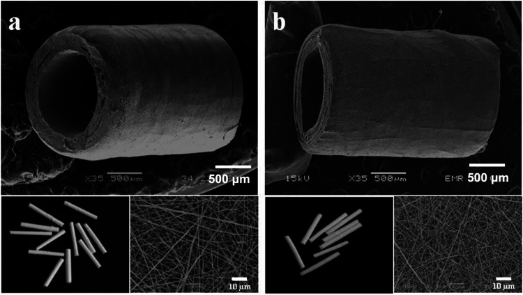
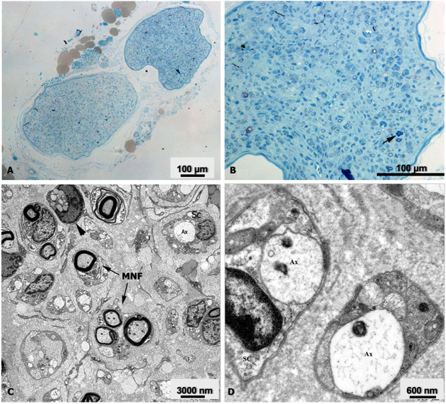
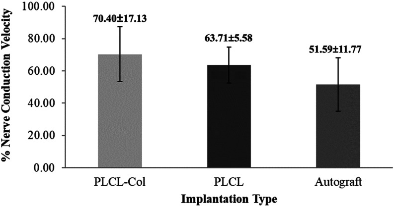
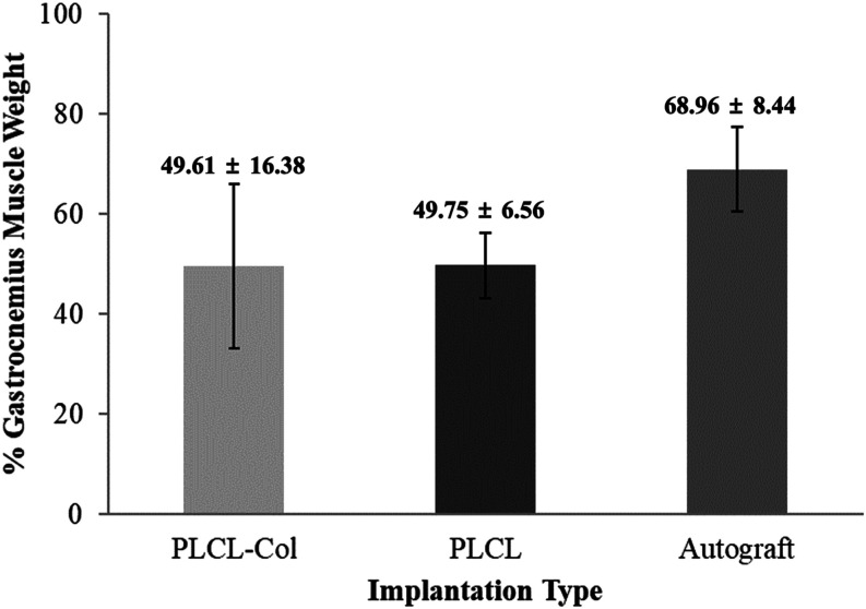
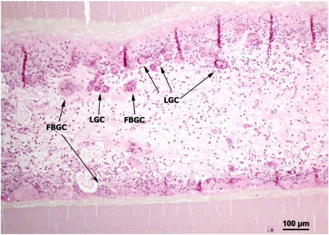
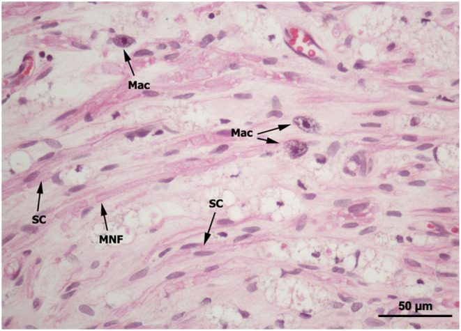
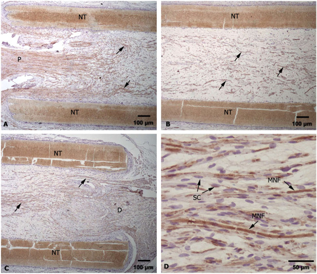
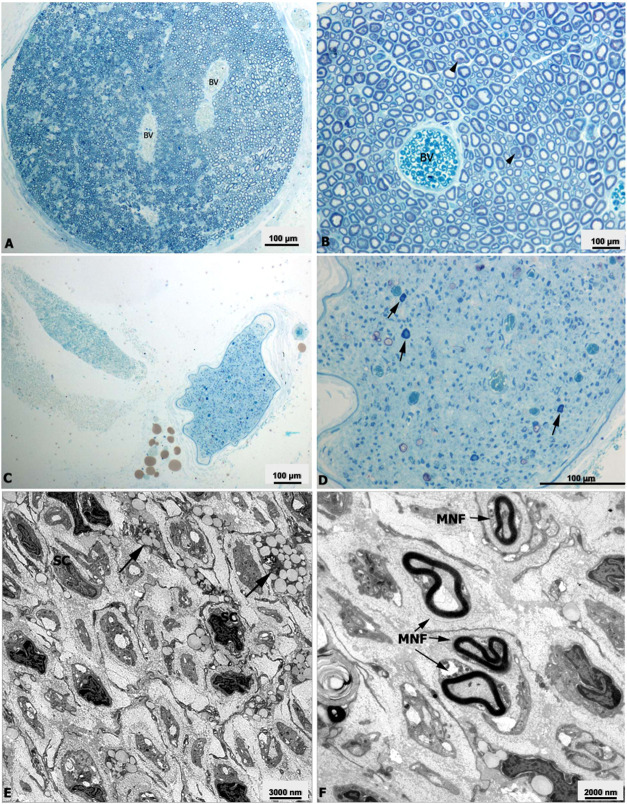
The present study provides trials of electrospun poly(l-lactide--caprolactone), PLCL, copolymer 67:33 mol %, and electrospun PLCL blend with a low loading of collagen (0.5% w/v), PLCL-Col, as a connecting porous biodegradable nerve conduit to repair 7 mm long segmentary tibial nerve lesions in rats compared with the standard autograft technique. The electrospun PLCL scaffolds reveal a matrix of fibers with a mean diameter of 476 ± 60 nm and an average pore size of 253 ± 5 nm. Blending collagen with the PLCL results in a comparatively denser matrix of fibers with a mean diameter of 417 ± 42 nm and a pore size of 244 ± 3 nm. For testing, a total of 30 male Wistar rats were divided into 3 groups of 10 and each group was subjected to a different nerve repair procedure for evaluation of nerve regeneration after reconstruction. Evaluation of nerve regeneration was compared in terms of the tibial functional index (TFI), nerve conduction velocity (NCV), gastrocnemius muscle weight (%GMW), and a histomorphometric study. After 12 weeks of implantation, there was evidence of nerve regeneration across the gap from the histomorphologic study. All parameters of nerve regeneration were observed in every animal of the study groups. Our results clearly showed that there are reinnervation and return of function in all groups, similarly to the autograft group. PLCL-Col showed better results than PLCL and autograft, which suggested that PLCL-Col porous conduits may serve as a scaffold for peripheral nerve regeneration.
本研究提供了电纺聚(L-丙交酯-己内酯)(PLCL)共聚物(67:33摩尔%),以及与低负载量胶原蛋白(0.5%w/v)混合的电纺PLCL(PLCL-Col),作为连接性多孔可生物降解神经导管,用于修复大鼠7毫米长的节段性胫神经损伤,并与标准自体移植技术进行比较。电纺PLCL支架呈现出纤维基质,平均直径为476±60纳米,平均孔径为253±5纳米。将胶原蛋白与PLCL混合会产生相对更致密的纤维基质,平均直径为417±42纳米,孔径为244±3纳米。为了进行测试,总共30只雄性Wistar大鼠被分为3组,每组10只,每组接受不同的神经修复程序,以评估重建后的神经再生情况。通过胫神经功能指数(TFI)、神经传导速度(NCV)、腓肠肌重量(%GMW)以及组织形态计量学研究来比较神经再生情况。植入12周后,组织形态学研究表明有神经再生跨越间隙的证据。研究组的每只动物都观察到了神经再生的所有参数。我们的结果清楚地表明,所有组都有神经再支配和功能恢复,与自体移植组相似。PLCL-Col的结果优于PLCL和自体移植组,这表明PLCL-Col多孔导管可作为周围神经再生的支架。