Kanahashi Toru, Imai Hirohiko, Otani Hiroki, Yamada Shigehito, Männer Jörg, Takakuwa Tetsuya
Human Health Science, Graduate School of Medicine, Kyoto University, Kyoto, Japan.
Innovation Research Center for Quantum Medicine, Gifu University School of Medicine, Gifu, Japan.
Cells Tissues Organs. 2025 Jun 19:1-17. doi: 10.1159/000546997.
While caudal foregut development in human fetuses has been outlined in previous research, the formation of its border region remains unclear. This study aimed to visualize the precise timeline of caudal foregut boundary formation.
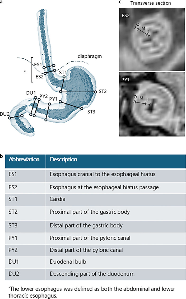
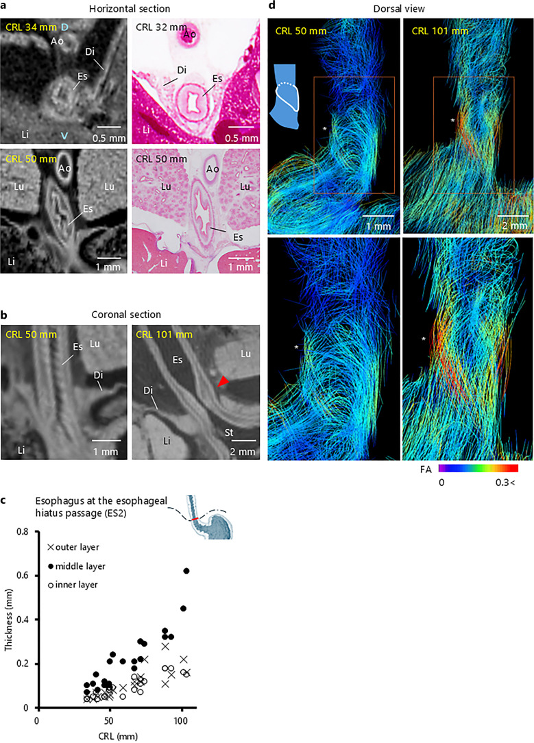
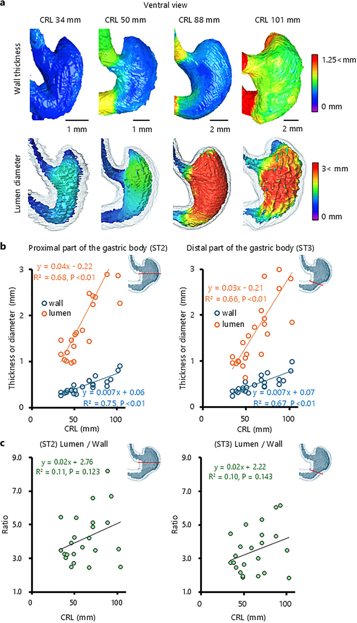
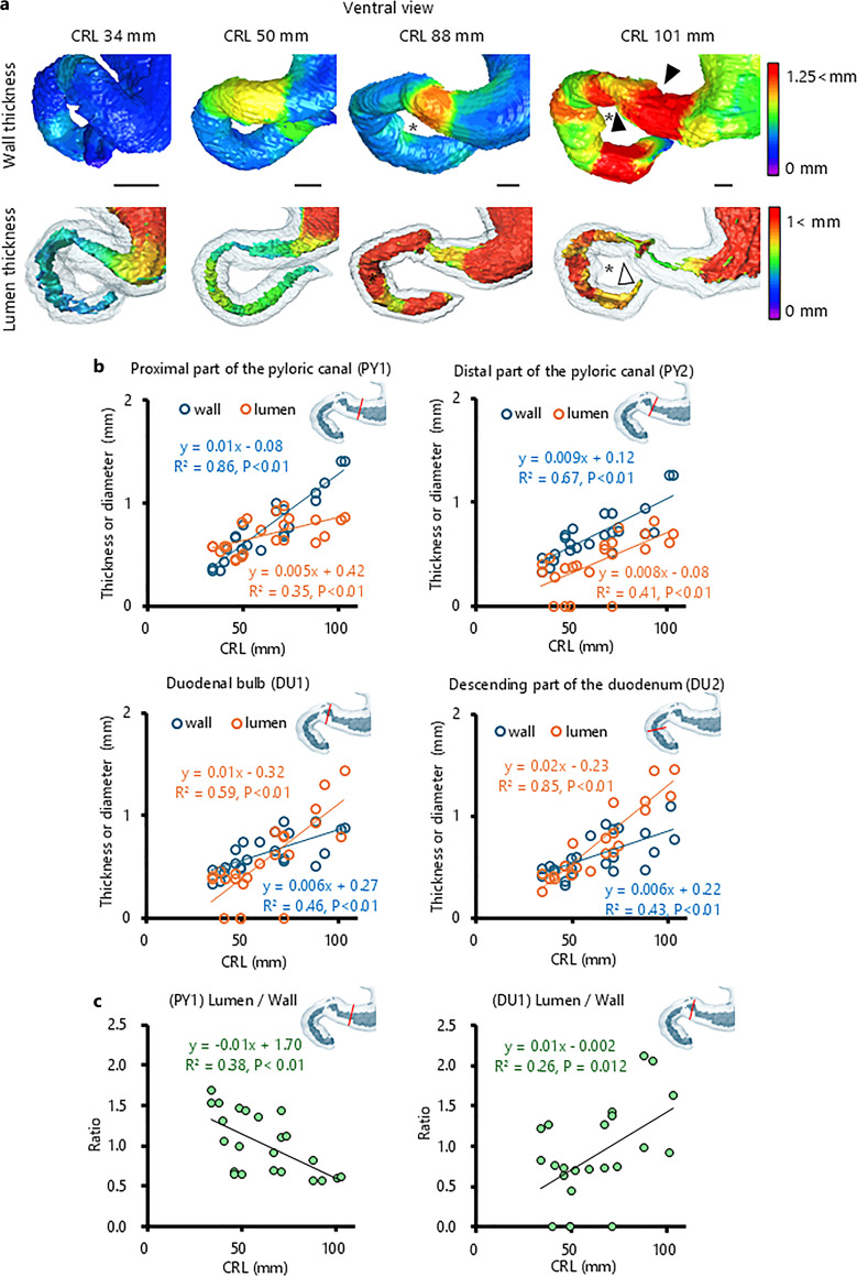
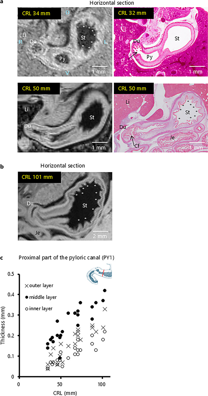
Three-dimensional images of the foregut from T1-weighted scans of 24 fetuses (crown-rump length [CRL]: 34-103 mm) were analyzed to measure the wall thickness and lumen diameter at nine specific sites. The internal structure in the border region was verified using histological sections and diffusion tensor imaging (DTI) tractography.
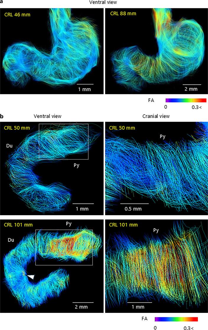
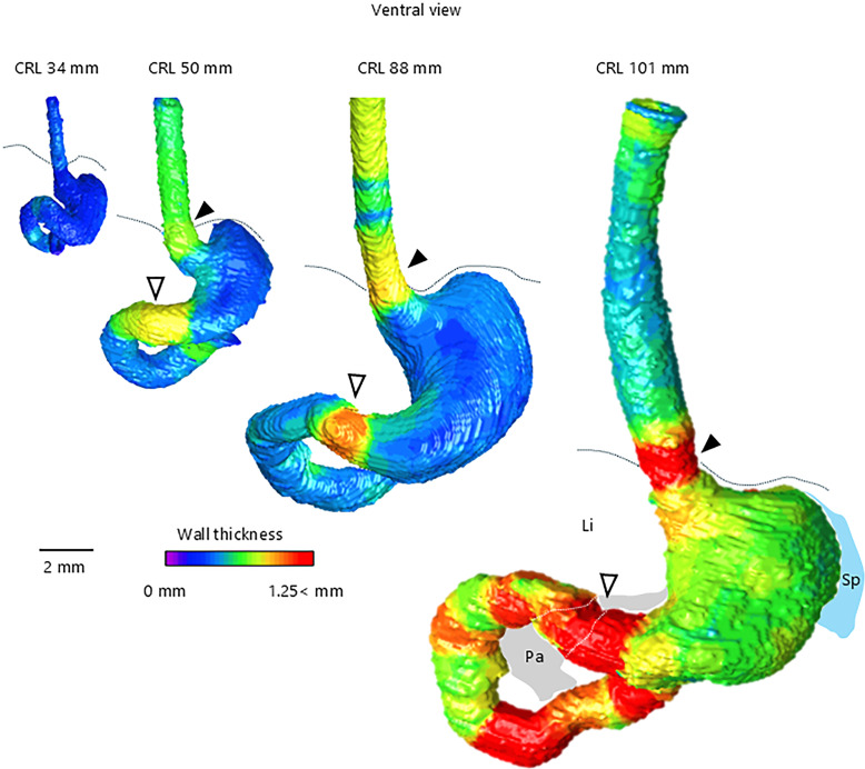
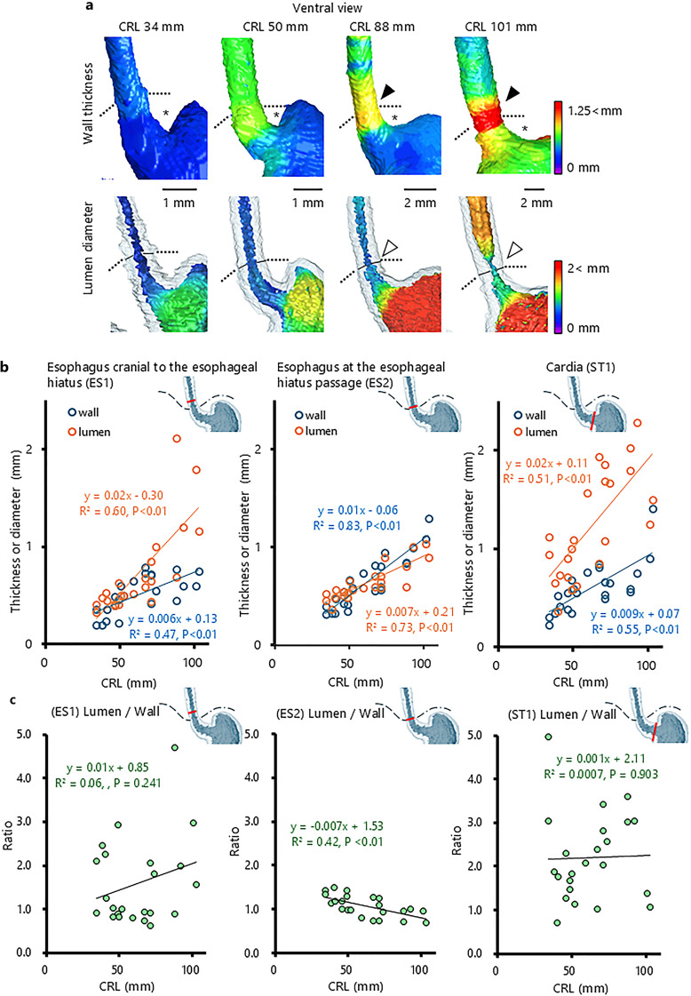
The lower esophageal and pyloric canal walls were thicker in samples with a CRL ≥50 mm. The esophageal wall at the esophageal hiatus, where the lower esophageal sphincter is located, was particularly thick in samples with a CRL ≥88 mm. Increased wall thickness at the esophageal hiatus and pyloric canal resulted in a narrower lumen. The pyloric canal lumen narrowed from its distal to proximal sections. The lumen diameter-to-wall thickness ratio at the esophageal hiatus and proximal pyloric was negatively correlated with CRL. The thickened esophageal wall at the esophageal hiatus had a thick submucosa, and all layers in the pyloric canal thickened with growth. DTI tractography revealed that the lower esophageal wall mainly comprised longitudinal fibers, whereas the pyloric canal wall consisted solely of circular fibers, with fractional anisotropy increasing with growth.
This study provides a comprehensive timeline of normal caudal foregut boundary formation during the early human fetal period, thereby improving the understanding of congenital foregut obstruction pathogenesis.
虽然先前的研究已经概述了人类胎儿前肠尾段的发育情况,但其边界区域的形成仍不清楚。本研究旨在可视化前肠尾段边界形成的精确时间线。
分析了24例胎儿(顶臀长[CRL]:34 - 103毫米)T1加权扫描的前肠三维图像,以测量九个特定部位的壁厚和管腔直径。使用组织学切片和扩散张量成像(DTI)纤维束成像验证边界区域的内部结构。
CRL≥50毫米的样本中,食管下段和幽门管的壁更厚。在食管裂孔处(食管下括约肌所在位置)的食管壁,CRL≥88毫米的样本中尤其厚。食管裂孔和幽门管处壁厚增加导致管腔变窄。幽门管腔从远端到近端逐渐变窄。食管裂孔和幽门近端的管腔直径与壁厚之比与CRL呈负相关。食管裂孔处增厚的食管壁有一层厚厚的黏膜下层,幽门管的所有层都随生长而增厚。DTI纤维束成像显示,食管下段壁主要由纵向纤维组成,而幽门管壁仅由环形纤维组成,分数各向异性随生长增加。
本研究提供了人类胎儿早期前肠尾段正常边界形成的全面时间线,从而增进了对先天性前肠梗阻发病机制的理解。