Świetlik Dariusz
Division of Biostatistics and Neural Networks, Medical University of Gdansk, Debinki 1 St., 80-211 Gdansk, Poland.
Biomedicines. 2025 Jun 9;13(6):1416. doi: 10.3390/biomedicines13061416.
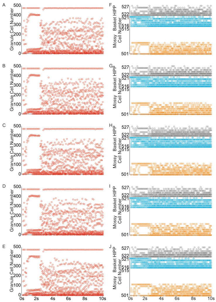
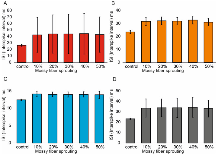
: A concussive head injury increases the likelihood of temporal lobe epilepsy through mechanisms that are not entirely understood. This study aimed to investigate how two key histopathological features shared by both TLE (temporal lobe epilepsy) and head injury-mossy fiber sprouting and hilar excitatory cell loss-contribute to the modulation of dentate gyrus excitability. : A computational approach was used to explore the impact of specific levels of mossy fiber sprouting and mossy cell loss, while avoiding the confounding effects of concurrent changes. The dentate gyrus model consists of 500 granule cells, 15 mossy cells, 6 basket cells and 6 hilar perforant path-associated cells. : My simulations demonstrate a correlation between the degree of mossy fiber sprouting and the number of spikes in dentate gyrus granule cells (correlations coefficient R = 0.95, < 0.0001) and other cells (correlations coefficient R = 0.99, < 0.0001). The mean values (standard deviation, SD) and 95% CI for granule cell activity in the control group and percentage 10-50% of mossy fiber sprouting groups are 376.4 (16.7) (95% CI, 374.9-377.8) vs. 463.5 (24.3) (95% CI, 461.4-465.6) vs. 514.8 (32.5) (95% CI, 511.9-517.6) vs. 555.0 (40.4) (95% CI, 551.5-558.6) vs. 633.4 (51.8) (95% CI, 628.8-637.9) vs. 701.7 (66.2) (95% CI, 695.9-707.5). The increase in mossy fiber sprouting was significantly statistically associated with an increase in granule cell activity ( < 0.01). The removal of mossy cells led to a reduction in excitability within the model network (for granule cells, correlations coefficient R = -0.40, < 0.0001). : These results are generally consistent with experimental observations, which indicate a high degree of mossy fiber sprouting in animals with a higher frequency of seizures. Whereas unlike the strong hyperexcitability effects induced by mossy fiber sprouting, the removal of mossy cells led to reduced granule cell responses to perforant path activation.
震荡性头部损伤会通过一些尚未完全明确的机制增加颞叶癫痫的发病可能性。本研究旨在探究颞叶癫痫(TLE)和头部损伤共有的两个关键组织病理学特征——苔藓纤维增生和门区兴奋性细胞丢失——如何调节齿状回的兴奋性。采用一种计算方法来探究特定程度的苔藓纤维增生和苔藓细胞丢失所产生的影响,同时避免并发变化带来的混淆效应。齿状回模型由500个颗粒细胞、15个苔藓细胞、6个篮状细胞和6个与门区穿通路径相关的细胞组成。我的模拟结果表明,苔藓纤维增生程度与齿状回颗粒细胞的放电次数之间存在相关性(相关系数R = 0.95,P < 0.0001),与其他细胞也存在相关性(相关系数R = 0.99,P < 0.0001)。对照组以及苔藓纤维增生比例为10% - 50%的各实验组中,颗粒细胞活动的平均值(标准差,SD)及95%置信区间分别为:376.4(16.7)(95% CI,374.9 - 377.8);463.5(24.3)(95% CI,461.4 - 465.6);514.8(32.5)(95% CI,511.9 - 517.6);555.0(40.4)(95% CI,551.5 - 558.6);633.4(51.8)(95% CI,628.8 - 637.9);701.7(66.2)(95% CI,695.9 - 707.5)。苔藓纤维增生的增加与颗粒细胞活动的增加在统计学上具有显著相关性(P < 0.01)。苔藓细胞的去除导致模型网络内的兴奋性降低(对于颗粒细胞,相关系数R = -0.40,P < 0.0001)。这些结果总体上与实验观察结果一致,实验观察表明癫痫发作频率较高的动物中存在高度的苔藓纤维增生。然而,与苔藓纤维增生所诱导的强烈的过度兴奋效应不同,苔藓细胞的去除导致颗粒细胞对穿通路径激活的反应减弱。