Li Xueqi, Li Xiangyu, Tong Lingjun, Hu Liqiao, Hong Yanfen, Zhou Ruoyu, Li Zonghong, Dong Ming, Hou Junjie, Xu Tao, Zhong Wen
Guangzhou Institute of Cancer Research, the Affiliated Cancer Hospital, Guangzhou Medical University Guangzhou China.
Guangzhou National Laboratory Guangzhou China.
J Extracell Biol. 2025 Jun 26;4(6):e70058. doi: 10.1002/jex2.70058. eCollection 2025 Jun.
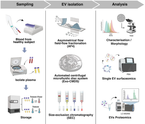
Extracellular vesicles (EVs) are increasingly recognized as promising disease biomarkers and therapeutic carriers. However, standardizing blood-derived EV isolation remains challenging due to the heterogeneity of EV populations and variability among isolation techniques. In this study, we systematically evaluated three distinct EV isolation methods, including asymmetrical flow field-flow fractionation (AF4), size-exclusion chromatography (SEC) and automated centrifugal microfluidic disc system combined with functionalized membranes (Exo-CMDS), to compare their efficiency in isolating EVs from both freshly frozen and freeze-thawed plasma samples. We utilized an integrative approach combining Proximity-dependent Barcoding Assay (PBA) for single-EV surface protein profiling, Liquid Chromatography-Mass Spectrometry (LC-MS/MS) for bulk proteomic analysis, along with transmission electron microscopy (TEM) and nanoparticle tracking analysis (NTA) to assess EV yield, morphology, surface protein expression and subpopulation diversity. Our results revealed significant differences in three EV isolation methods. AF4 is particularly enriched for EV subpopulations expressing high levels of classical tetraspanins (e.g., CD81, CD9 and CD151), and single-pass membrane proteins (e.g., ITGA4 and ITAGB1). Exo-CMDS demonstrated the highest reproducibility across samples, isolating specific EV subpopulations enriched in markers like CD5. SEC provided the highest yield but co-isolated significant amounts of non-vesicular particles, including lipoproteins. The findings contribute valuable insights toward standardized and reliable EV isolation practices for research and clinical applications.
细胞外囊泡(EVs)越来越被认为是有前景的疾病生物标志物和治疗载体。然而,由于EV群体的异质性和分离技术之间的差异,标准化血液来源的EV分离仍然具有挑战性。在本研究中,我们系统地评估了三种不同的EV分离方法,包括不对称流场-流分级分离(AF4)、尺寸排阻色谱(SEC)和结合功能化膜的自动离心微流盘系统(Exo-CMDS),以比较它们从新鲜冷冻和冻融血浆样本中分离EVs的效率。我们采用了一种综合方法,结合用于单个EV表面蛋白分析的邻近依赖性条形码分析(PBA)、用于整体蛋白质组分析的液相色谱-质谱联用(LC-MS/MS),以及透射电子显微镜(TEM)和纳米颗粒跟踪分析(NTA)来评估EV产量、形态、表面蛋白表达和亚群多样性。我们的结果揭示了三种EV分离方法之间的显著差异。AF4特别富集表达高水平经典四跨膜蛋白(如CD81、CD9和CD151)和单次跨膜蛋白(如ITGA4和ITAGB1)的EV亚群。Exo-CMDS在不同样本中表现出最高的重现性,分离出富含CD5等标志物的特定EV亚群。SEC产量最高,但共分离出大量非囊泡颗粒,包括脂蛋白。这些发现为研究和临床应用中标准化和可靠的EV分离实践提供了有价值的见解。